


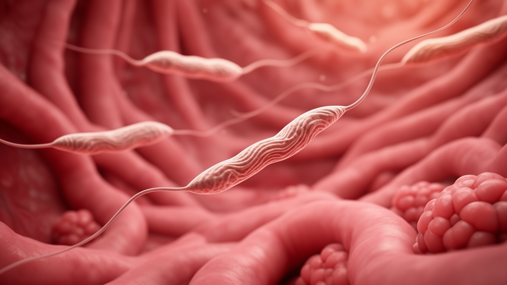
Human African trypanosomiasis, better known as sleeping sickness, has long been understood as a disease of the blood, the immune system, and ultimately the brain. Caused by the single-celled parasite Trypanosoma brucei and transmitted by tsetse flies, the infection is notorious for the fever, wasting, and neurological decline that define its progression. Yet a new study published in PLOS Pathogens suggests that the damage inflicted by this parasite extends into a region of the body that has received far less attention: the female reproductive tract. Using a mouse model of infection, researchers found that T. brucei physically invades the lining of the uterus, remodels its immune landscape, and sets off a cascade of hormonal and metabolic disruption that ultimately halts the reproductive cycle.
The research team, led by Olivia M. Shorthouse and Juan F. Quintana, set out to answer a deceptively simple question. Reproductive dysfunction has been reported anecdotally in both human patients and experimental animals infected with T. brucei, but nobody knew whether the parasites actually entered the female reproductive tract or whether the observed problems were merely secondary effects of systemic illness. By tracking parasite localisation during both acute and chronic stages of infection in mice, the investigators demonstrated that the organisms take up residence in the endometrium, the specialised mucosal lining of the uterus. This direct infiltration of reproductive tissue places the parasite in intimate contact with the immune cells and hormone-responsive epithelium that govern fertility.
What the parasites do once they arrive proved to be as important as where they settle. The study revealed a progressive and multi-layered deterioration of reproductive health as infection moved from its acute phase into chronicity. Chronically infected mice lost substantial fat mass, a sign of the metabolic wasting characteristic of trypanosome disease. Their oestrous cycles, the rodent equivalent of the menstrual cycle, became disrupted and eventually arrested. The uterus and ovaries themselves shrank, showing measurable atrophy. Perhaps most strikingly, the researchers documented extensive transcriptional dysregulation across the hypothalamic-pituitary-gonadal axis, the hormonal control circuit that links the brain to the gonads and orchestrates fertility from the top down.
The hypothalamic-pituitary-gonadal axis is a delicate three-tiered system. The hypothalamus, a small region at the base of the brain, releases gonadotropin-releasing hormone in pulses that instruct the pituitary gland to secrete luteinising hormone and follicle-stimulating hormone. These hormones, in turn, act on the ovaries to drive follicle development, ovulation, and the production of oestrogen and progesterone. The new findings show that chronic T. brucei infection distorts gene expression at every level of this hierarchy, implying that the parasite does not merely damage the uterus locally but interferes with the central neuroendocrine commands that regulate it. This systemic rewiring of reproductive control represents a form of pathology that has rarely been characterised in parasitic infection.
Immune remodelling within the uterus emerged as a central feature of the disease process. In both acute and chronic infection, the researchers observed infiltration of T cells into uterine tissue and activation of pro-inflammatory myeloid cells, the innate immune population that includes monocytes, macrophages, and their inflammatory descendants. Beyond the uterus itself, the team detected broader type 1 inflammatory signatures spreading across reproductive tissues and components of the hypothalamic-pituitary-gonadal axis. Type 1 inflammation, typically driven by interferon signalling and associated with defence against intracellular pathogens, is not normally the dominant immune tone of the reproductive tract, which tends to favour tolerance and controlled responses. Its expansion here suggests that the infection fundamentally reprograms the immunological identity of the female reproductive organs.
The ovaries told their own story of damage. Examination of ovarian pathology revealed follicular degeneration, meaning the structures that house and nurture developing eggs were deteriorating. The researchers also counted a reduction in corpora lutea, the transient endocrine structures that form after ovulation and produce the progesterone needed to support early pregnancy. Fewer corpora lutea indicate fewer successful ovulations, a direct readout of impaired fertility. Alongside these structural changes, alterations to steroidogenic pathways were detected, pointing to disruption of the enzymatic machinery that converts cholesterol into the steroid hormones essential for cyclicity and reproduction.
One of the most compelling experiments in the study involved a therapeutic intervention designed to disentangle two competing explanations. If the uterine damage were driven purely by falling oestrogen levels, then restoring oestrogen signalling should rescue the tissue. The researchers treated infected mice with tamoxifen, a selective oestrogen receptor modulator widely known for its use in breast cancer therapy. The results were revealing but partial. Tamoxifen restored uterine morphology and prevented the arrest of the oestrous cycle, demonstrating that endocrine dysfunction is indeed a major driver of the structural collapse of the uterus. However, the hormone treatment did not reverse the infection-induced remodelling of the uterine immune environment. The inflammatory cell populations and their activation states persisted despite hormonal rescue.
This dissociation carries a significant conceptual message: endocrine dysfunction and infection-driven inflammation are two distinct processes, each contributing to reproductive pathology through separate routes. Hormonal collapse explains the loss of normal uterine architecture and cyclicity, but the immune reprogramming is a direct consequence of the infection itself and cannot be corrected simply by fixing the hormonal signal. For patients, this implies that treating the hormonal consequences of chronic parasitic disease may not be sufficient to restore full reproductive health unless the underlying inflammatory drive, and ideally the infection itself, is also addressed. It also raises the possibility that immune-mediated damage to the reproductive tract could outlast or operate independently of the visible endocrine symptoms.
The broader significance of the work lies in what it says about systemic infection as a whole. The female reproductive tract is an immunologically dynamic organ that must balance defence against pathogens with the tolerance required for successful reproduction. The new findings identify it as a major target of T. brucei infection and demonstrate how a chronic parasitic disease can disrupt reproductive physiology through a combination of immune, endocrine, and metabolic pathways acting in parallel. Fat wasting, hypothalamic dysregulation, ovarian degeneration, and uterine inflammation are not isolated phenomena but interconnected consequences of a systemic infection that reaches into every tier of reproductive control. The authors suggest that this interplay between metabolism, immunity, and endocrinology may be relevant well beyond trypanosomiasis.
Indeed, the study closes with a methodological warning for the wider research community. Because the female reproductive tract has been so frequently overlooked in models of systemic inflammation, the effects of other infectious and inflammatory diseases on fertility and reproductive endocrinology may be underappreciated. The researchers argue that future studies of systemic inflammatory conditions should specifically assess the female reproductive tract rather than assuming it is a passive bystander. For sleeping sickness, a disease that affects tens of thousands of people in sub-Saharan Africa, the findings open a new dimension of clinical concern and a new set of questions about how parasitic infection shapes long-term reproductive health in survivors. They also provide a framework for investigating whether other chronic infections quietly inflict similar damage on the reproductive system.
Subject of Research: Trypanosoma brucei infection of the female reproductive tract and its effects on uterine immunity and reproductive hormone regulation
Article Title: Trypanosoma brucei infection remodels the uterine immune environment and drives neuroendocrine dysfunction
Article References: Shorthouse, O. M., Barnes, C., Colombo, S. A. P., Costa, J., Wonsbek, K., Mohon, A., MacDonald, A. S., Mann, E., Costain, A. H., & Quintana, J. F. (2026). Trypanosoma brucei infection remodels the uterine immune environment and drives neuroendocrine dysfunction. PLOS Pathogens, 22(10), e1014667. https://doi.org/10.1371/journal.ppat.1014667
Image Credits: AI Generated
DOI: 10.1371/journal.ppat.1014667
Keywords: Trypanosoma brucei, sleeping sickness, uterus, endometrium, reproductive immunology, hypothalamic-pituitary-gonadal axis, oestrous cycle, ovarian pathology, tamoxifen, inflammation, parasitology, PLOS Pathogens
Cite Scienmag News
Kristina Jarvis. (October 9, 2026). Sleeping Sickness Parasite Invades the Uterus and Disrupts Reproductive Hormones. Scienmag. https://scienmag.com/sleeping-sickness-parasite-invades-the-uterus-and-disrupts-reproductive-hormones/
Kristina Jarvis. "Sleeping Sickness Parasite Invades the Uterus and Disrupts Reproductive Hormones." Scienmag, 9 October 2026, https://scienmag.com/sleeping-sickness-parasite-invades-the-uterus-and-disrupts-reproductive-hormones/. Accessed 9 October 2026.
Kristina Jarvis. "Sleeping Sickness Parasite Invades the Uterus and Disrupts Reproductive Hormones." Scienmag. October 9, 2026. https://scienmag.com/sleeping-sickness-parasite-invades-the-uterus-and-disrupts-reproductive-hormones/


