


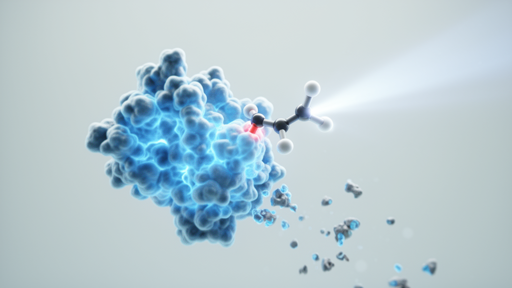
Chemists have long dreamed of drugs that work only where and when doctors want them to. A new study published in Nature Communications brings that vision closer, describing a class of ruthenium-based molecules that can be switched on with light to destroy specific proteins inside living cells. The compounds, called RuPHOTACs, short for Ruthenium-based PHOToActivated Chimeras, combine the power of targeted protein degradation with the precision of photochemistry, and the researchers report that they show the highest selectivity for light versus dark activation yet achieved in this emerging field.
To understand why this matters, it helps to start with PROTACs, or PROteolysis TArgeting Chimeras. These bifunctional molecules are the flagship technology of targeted protein degradation, one of the most exciting areas of modern drug discovery. A PROTAC has two business ends: one binds a protein that scientists want eliminated, and the other recruits an E3 ubiquitin ligase, the cellular machinery that tags unwanted proteins for destruction by the proteasome. By physically dragging the target protein into contact with the ligase, a PROTAC catalyzes the formation of a protein-protein interaction that would never happen naturally, triggering the cell’s own waste-disposal system to chew up the target. Unlike conventional inhibitors, which merely block a protein’s activity and must be present at high concentrations to keep it suppressed, degraders can eliminate the protein altogether and work catalytically, so a single molecule can destroy many copies of its target.
The problem is that this power comes with a loss of control. Because PROTACs act anywhere they reach in the body, they can degrade their target proteins in healthy tissues as well as diseased ones, producing side effects that are difficult to predict. The field has therefore been searching for ways to impose spatiotemporal control, meaning the ability to restrict drug activity to a specific place at a specific time. Light is the most attractive remote trigger for this purpose, since it can be focused on a tissue with exquisite precision and switched on and off at will. Photocaging strategies, in which a light-sensitive chemical group masks a drug’s activity until illumination releases it, have been applied to PROTACs before, but earlier systems suffered from weak contrast between the dark and illuminated states, or required high-energy ultraviolet light that damages cells and penetrates poorly into tissue.
The team behind the new work, led by Dmytro Havrylyuk, David K. Heidary and Edith C. Glazer at North Carolina State University, together with colleagues at the University of Kentucky, took a different approach by building the light switch out of a metal. Their RuPHOTACs use ruthenium(II) coordination complexes as photocages wrapped around the PROTAC scaffold. Ruthenium complexes have a distinctive photochemistry: when they absorb light, they can undergo ligand photodissociation, cleanly releasing the molecule attached to the metal. Crucially, the absorption properties of these complexes can be tuned so that the release is driven by low-energy red light rather than damaging ultraviolet wavelengths. Red light penetrates deeper into biological tissue and is far gentler on living cells, which is why the authors emphasize that their systems are optimized for biological applications rather than merely demonstrated in a cuvette.
To prove the concept, the researchers targeted bromodomain-containing proteins, a family of epigenetic regulators that read acetylation marks on histones and help control gene expression. These proteins, particularly members of the BET family, are major targets in cancer research because many tumors depend on them to drive the expression of growth-promoting genes. In the dark, the ruthenium cage keeps the RuPHOTAC inert; when illuminated with red light, the cage releases the active degrader, which then recruits the E3 ligase and drags the bromodomain protein to the proteasome. The team showed that the compounds strongly reduced not only the levels of the bromodomain targets themselves but also the abundance of two important downstream proteins, c-MYC and PIM1, both of which are transcriptional targets of BET proteins and both of which are heavily implicated in cancer progression. Knocking down c-MYC in particular is a longstanding goal in oncology, since the protein has been notoriously difficult to drug with conventional small molecules.
The quantitative performance of the new compounds is what sets the study apart. The authors report that the RuPHOTACs achieve the highest selectivity for activation in the light versus the dark reported for photocontrolled degraders, meaning that in the absence of light the molecules are essentially silent, while a brief illumination unleashes full degrading activity. They also show improved potency against the target proteins compared with prior systems, so less compound is needed to achieve the same degree of degradation. Most strikingly, the increased efficacy extends to animal work: using low-energy red light, the team demonstrated increased efficacy in vivo, a critical benchmark because many photochemical technologies that shine in cell culture fail when confronted with the optical and biological complexity of a living organism.
Measuring protein degradation sounds simple but is surprisingly tricky, and the study introduces an elegant solution to a chronic measurement problem. Traditional assays often rely on antibodies or fluorescent tags that report total protein abundance, but those signals conflate two opposing processes: the destruction of existing protein molecules and the continuous production of new ones. If a drug slows protein synthesis as a side effect, or if the cell ramps up production to compensate for degradation, the apparent degradation rate becomes distorted. To disentangle these effects, the researchers created fusion proteins in which the target of interest is fused to Dendra2, a photoconvertible fluorescent protein. Dendra2 can be switched from a green to a red fluorescent state by light, allowing the researchers to mark a defined cohort of pre-existing protein molecules and then watch that cohort disappear over time as the PROTAC does its work.
This bifunctional reporter system offers several advantages over previously described PROTAC reporters. Because the photoconverted molecules are already synthesized, any change in the red fluorescence signal reflects true degradation rather than changes in new protein production. At the same time, the green signal tracks newly synthesized protein, so the researchers can monitor both destruction and replenishment simultaneously in the same living cell. Importantly, the approach does not perturb translation, proteostasis, or cell health, meaning the measurements reflect normal cellular physiology rather than artifacts introduced by the measurement itself. The authors argue that this makes the system superior to prior reporter strategies for live-cell analysis of protein degradation, and it could become a standard tool for the broader targeted degradation community, which has long struggled with assay artifacts.
The study also carries a broader message about the role of metals in chemical biology. Ruthenium is not a component of any natural biomolecule, and metal complexes are often viewed with suspicion in drug development because of concerns about stability and toxicity. Yet the work demonstrates that incorporating a metal component within an organic PROTAC confers multiple concrete advantages: a clean and efficient photorelease mechanism, absorption profiles compatible with low-energy red light, and tunable photophysical properties that organic photocages struggle to match. The researchers suggest that this design philosophy, using coordination chemistry as an engineering platform for light-controlled drugs, could extend well beyond the specific bromodomain targets validated here, potentially allowing any PROTAC scaffold to be fitted with a ruthenium switch.
Caveats remain, as they always do at this stage of translation. The work was supported by the National Institutes of Health and the Kentucky Cabinet for Health and Family Services, and the authors declare no competing interests, but moving from validated compounds in cells and animal models toward clinical candidates will require extensive safety evaluation of the ruthenium payloads, optimization of light delivery to internal organs, and pharmacokinetic studies that the current paper does not address. The article itself was shared early as a peer-reviewed accepted manuscript subject to further editorial processing. Even so, the combination of near-total dark-state silence, red-light activation, in vivo efficacy, and a genuinely improved measurement technology makes RuPHOTACs one of the most complete demonstrations to date that protein degradation can be placed under remote optical control. For a field racing to make degraders safer and more precise, that is a result worth watching closely.
Subject of Research: Light-activated ruthenium photocaged PROTACs for spatiotemporally controlled targeted protein degradation
Article Title: RuPHOTACs provide photocontrol over protein degradation with optimized properties for biological applications
Article References: Havrylyuk, D., LaMore, A. P., Al-Hamaly, M. A., Blackburn, J. S., Heidary, D. K., & Glazer, E. C. (2026). RuPHOTACs provide photocontrol over protein degradation with optimized properties for biological applications. Nature Communications. https://doi.org/10.1038/s41467-026-78110-x
Image Credits: AI Generated
DOI: 10.1038/s41467-026-78110-x
Keywords: targeted protein degradation, PROTACs, RuPHOTACs, ruthenium photocages, photochemistry, E3 ubiquitin ligase, bromodomain proteins, c-MYC, Dendra2 reporter, red light activation, chemical biology, cancer research
Cite Scienmag News
Ophelia Keating. (October 9, 2026). Light-Activated Molecular Degraders Put Protein Destruction Under Remote Control. Scienmag. https://scienmag.com/light-activated-molecular-degraders-put-protein-destruction-under-remote-control/
Ophelia Keating. "Light-Activated Molecular Degraders Put Protein Destruction Under Remote Control." Scienmag, 9 October 2026, https://scienmag.com/light-activated-molecular-degraders-put-protein-destruction-under-remote-control/. Accessed 9 October 2026.
Ophelia Keating. "Light-Activated Molecular Degraders Put Protein Destruction Under Remote Control." Scienmag. October 9, 2026. https://scienmag.com/light-activated-molecular-degraders-put-protein-destruction-under-remote-control/


