

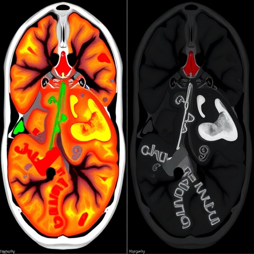
A groundbreaking tool is set to democratize the intricate world of medical image analysis by stripping away the need for advanced programming skills. Detailed in a new protocol published in Nature Protocols, researchers have unveiled CODAvision, a graphical user interface that guides scientists through the notoriously complex process of training deep learning algorithms for semantic segmentation. This development promises to place the power of artificial intelligence directly into the hands of biologists, clinicians, and pathologists who have long been barred from building custom image analysis models by unwieldy code and arcane parameter tuning.
At its core, semantic segmentation is the pixel-level assignment of identity to every structure within an image—distinguishing a cancerous nucleus from a healthy stromal cell, or a ventricle from surrounding brain tissue. Historically, generating the labeled datasets and training the convolutional neural networks that perform this task has demanded fluency in Python, mastery of deep learning frameworks like TensorFlow or PyTorch, and a deep understanding of data augmentation strategies. CODAvision collapses these hurdles into a point-and-click ecosystem, automating the configuration of model architectures, learning rates, and loss functions while providing real-time feedback on training progress.
The protocol’s technical backbone is the CODA (COmputational Digital Anatomy) algorithm, which has been retrofitted with a user-friendly interface. Unlike off-the-shelf segmentation solutions that force users into rigid, pre-trained categories, CODAvision allows for the rapid creation of highly bespoke models. A researcher studying metastatic burden in mouse models, for example, can upload a handful of annotated histology slides, visually inspect the network’s predictions against ground truth, and iteratively refine the model’s performance through an intuitive validation dashboard. The system automatically handles image normalization, tiling of gigapixel whole-slide images, and the augmentation of training data with rotations, flips, and elastic deformations that prevent the model from overfitting to a single staining batch or scanner type.
One of the most compelling aspects of the CODAvision workflow is its radical cross-modality versatility. The team demonstrates robust performance not only on conventional hematoxylin and eosin stained sections but also on magnetic resonance imaging (MRI), computed tomography (CT), and even the deconvolution of spot-based spatial transcriptomics datasets. This means a single interface can be used to quantify tumor volume from an MRI scan in the morning and to map the spatial distribution of gene expression within a tissue section in the afternoon, all without writing a single line of code. Under the hood, the system leverages a modified U-Net architecture with attention gates that learn to suppress irrelevant background regions, a design choice proven to enhance boundary delineation across highly heterogeneous image textures.
The protocol outlines a set of best practices that read like a masterclass in medical image analysis. It emphasizes the importance of inter-rater variability and instructs users on how to generate consensus annotations from multiple experts using the Stevens’ method, a statistical approach that weights annotator reliability. For model evaluation, CODAvision computes an exhaustive panel of metrics including Dice similarity coefficient, Hausdorff distance, and average surface distance, presenting them in a generated comprehensive report that can be directly cited in publications. This transparency sidesteps the common pitfall of relying solely on the Dice score, which can be misleading when segmenting small or thin structures.
A particularly innovative demonstration lies in the application to metastatic burden quantification in in vivo models. The protocol shows how CODAvision can be trained to detect micro-metastases in lymph nodes and visceral organs with a sensitivity that rivals manual counting by a trained pathologist, but at a throughput impossible for a human to sustain. The deep learning model learns to suppress normal tissue architectures while highlighting clusters of malignant cells, automatically outputting a spatial heatmap of metastatic probability that can be overlaid onto the original image. This capability has immediate implications for preclinical drug development, where the ability to precisely measure treatment response across entire animal cohorts can significantly accelerate compound triage.
Equally transformative is the tool’s integration with spatial omics. Spot-based spatial transcriptomics platforms like Visium generate tissue images where each spot corresponds to an array of gene expression values. By applying CODAvision’s semantic segmentation to these images, researchers can deconvolve the cellular composition underlying each spot, transforming a coarse expression map into a high-resolution atlas of cell types. The protocol details how a model trained to recognize histological features can infer the local mixture of epithelial, immune, and stromal components, enabling spatially aware differential expression analysis that is grounded in anatomical reality rather than blind clustering.
By converting the entire deep learning pipeline into a graphical experience, CODAvision fundamentally redefines the division of labor between computational and experimental scientists. Anatomists and biologists can now directly translate their domain expertise into robust, publication-grade quantitative outputs, while computational teams can focus on novel architecture design rather than the repetitive fine-tuning of standard models. The Nature Protocols paper serves as both a technical manual and a manifesto, envisioning a future where custom AI segmentation models become as routine a tool in the image analysis arsenal as the western blot quantification software is in molecular biology. The protocol is poised to unleash a wave of discoveries hidden within the terabytes of medical images generated daily, finally matching the power of deep learning to the questions that only domain experts know how to ask.
Subject of Research: A graphical user interface-based protocol for automatic semantic segmentation of medical images using the CODAvision deep learning algorithm.
Article Title: No-Code Deep Learning Brings Medical Image Segmentation to the Masses
Article References:
Matos-Romero, V., Gómez-Becerril, J., Forjaz, A. et al. CODAvision: best practices and a user-friendly interface for rapid, customizable segmentation of medical images.
Nat Protoc (2026). https://doi.org/10.1038/s41596-026-01404-3
Image Credits: AI Generated
DOI: https://doi.org/10.1038/s41596-026-01404-3
Keywords: semantic segmentation, deep learning, medical image analysis, graphical user interface, CODAvision, histology, magnetic resonance imaging, computed tomography, spatial transcriptomics, no-code AI


