


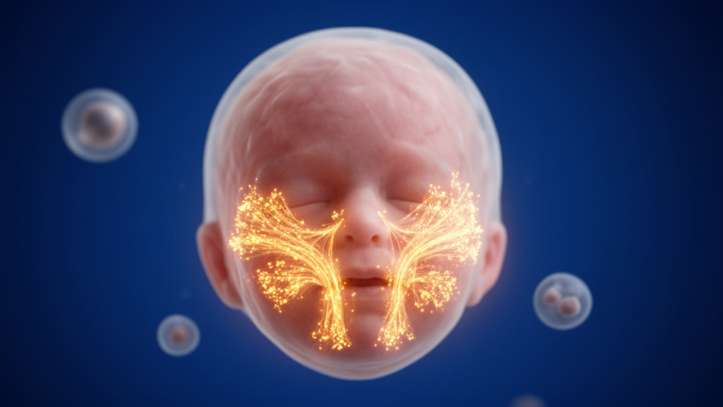
Why does every human face look different, and where in the developing embryo is that difference actually written? A large international team has now produced the most detailed answer yet, publishing in Nature Genetics a multimodal atlas of the human craniofacial region that charts, cell by cell and week by week, the molecular machinery that turns a featureless embryonic prominence into a nose, a jaw and a pair of cheekbones. By combining single-cell transcriptomics, chromatin accessibility profiling and high-resolution spatial transcriptomics across embryonic weeks six to eleven, the researchers captured fifty-six distinct cell states and traced how genetic variants scattered across the noncoding genome reach into these cells to shape the face each of us carries through life.
The biological starting point is well established in outline. The human face arises primarily from cranial neural crest cells, a migratory embryonic population that pours into the facial prominences and differentiates into ectomesenchyme, the tissue that will generate cartilage, bone, dermis, perivascular cells and more. Epithelial signaling centers and the forebrain then induce skeletogenic condensations, which form the cartilaginous chondrocranium, a template that helps define facial dimensions and jaw shape before it either ossifies directly or guides the deposition of dermal bone. What has remained stubbornly unclear is which specific cell states act as the main conduits through which the genome influences adult facial variation, particularly because the human face differs so dramatically from those of laboratory model organisms.
To close that gap, the team dissected embryonic craniofacial tissue under a stereo microscope into distinct regions, including the frontonasal, maxillary and mandibular prominences, and processed thirty single-cell RNA sequencing samples from eleven embryos alongside twelve paired single-nucleus RNA and ATAC sequencing samples from four individuals. The paired measurements allowed the researchers to connect gene expression to the chromatin landscape that regulates it, while microcomputed tomography of fixed embryos documented the changing skeletal architecture. The resulting dataset captured predominantly ectomesenchyme derivatives, including cartilage, bone, pericytes, meninges, dermal fibroblasts and tenocytes, which the team annotated into seventeen coarse-grained and fifty-six fine-grained cell clusters, each carrying its own signature of differentially accessible chromatin regions.
A key technical challenge was that mesenchymal subpopulations showed noisy, overlapping gene expression profiles, apparently because cells sampled from different anatomical regions carried position-dependent transcriptional programs. The researchers solved this with spatial transcriptomics, using Stereo-seq on frontal sections from week 6.5 embryos to build a high-resolution map of where each cell type actually sits within the growing face. They then applied Moran’s I autocorrelation statistics, a method that asks whether neighboring locations tend to share similar gene expression values, comparing spatial autocorrelation with transcriptional autocorrelation to distinguish genes that are location-specific from those that are merely cell-type-specific. This analysis yielded 396 so-called spatial genes, a molecular coordinate system for the embryonic face.
The spatial gene list proved strikingly enriched for transcription factor activity and, tellingly, for craniofacial disease phenotypes. Hierarchical clustering revealed nine modules of coexpressed transcription factors marking distinct facial domains, including the predermis, forming eyelids, nasal region, eye region, dental region, lower face and submandibular region. Binding sites for these spatial transcription factors were significantly enriched in cell-type-specific open chromatin regions, and enhancer-driven gene regulatory network inference confirmed widespread activity of these spatial networks across mesenchymal clusters. Known patterning regulators such as ALX1 and DLX5 showed region-specific, pseudotime-associated expression along trajectories of mesenchymal differentiation, suggesting that each facial region deploys its own dedicated module of transcription factors to direct site-specific differentiation.
Because most genetic variants associated with facial traits sit outside protein-coding regions, enhancers are the presumed vehicles through which the genome sculpts the face. The team assembled a map of roughly 50,000 candidate cis-regulatory elements whose chromatin accessibility correlates with nearby gene expression, and overlapped these with published facial enhancer datasets, cap analysis of gene expression data and H3K27ac enhancer marks. One standout candidate was an enhancer roughly twenty kilobases upstream of PAX1, a gene responsible for otofaciocervical syndrome, which carries a genome-wide significant variant associated with nasal width and shows cell-type-specific accessibility in early mesenchymal progenitors and dermal mesenchyme. A 276-base-pair segment of this enhancer is 94 percent conserved between mouse and human.
To test whether this conserved sequence matters in vivo, the researchers used CRISPR-based genome editing to delete it in mice. The edited animals consistently produced offspring with smaller bodies, shorter heads and apparent calvarial defects, including holes in the skull vault, while lacking other characteristic features of full Pax1 knockouts such as scoliosis or a kinked tail. The result provides experimental evidence that a single conserved enhancer identified in the human atlas is functionally important for mammalian skeletal morphology, though the authors caution that further work is needed to confirm that the deleted sequence directly interacts with PAX1 rather than acting through another mechanism.
Overlaying the atlas with a large facial genome-wide association study covering more than sixty facial traits revealed a clear temporal logic. Mesenchymal progenitor cells were significantly enriched for variants affecting broad, global facial dimensions, while more localized traits were associated with progressively more mature cell types, indicating that the influence of any given cell state on facial shape diminishes as development proceeds. The earliest mesenchymal progenitors, including a SOX11-positive cluster enriched at week 6.5, showed the strongest correspondence between where their genes were expressed and where nearby variants exerted their effects, particularly among the 396 spatial genes. Region-specific enhancers were linked to more than one hundred of these spatial genes, including an ALX1-linked enhancer eighty kilobases upstream that shows nose-specific accessibility and carries variants affecting the upper face. The authors conclude that variants within enhancers targeting key developmental genes are likely hotspots for generating facial diversity, and that multiple enhancers with distinct regional activation profiles can compositely establish a single gene’s expression domain, as illustrated for MSX1, a gene tied to cleft palate, oligodontia and Wolf-Hirschhorn syndrome.
Not every association followed the mesenchymal story. When the researchers examined GWAS data for orofacial clefts, epithelial cells, not mesenchyme, showed the strongest enrichment. They identified an epithelial subpopulation expressing the known cleft-related genes IRF6 and GRHL3, which act together in a circuit essential for timely periderm differentiation during palate formation, and pinpointed a single enhancer linked simultaneously to IRF6, LAMB3 and MIR205HG, with accessibility restricted to epithelial cells and overlapping known cleft-associated variants. Separately, signaling analysis predicted that peripheral neurons communicate with facial mesenchyme through Neuregulin and Pleiotrophin pathways, and mouse embryos lacking cranial sensory ganglia due to Neurogenin 1 knockout showed significant alterations in maxilla and alisphenoid shape, suggesting that nerves fine-tune the facial skeleton during embryonic development.
Together, the atlas reframes facial individuality as a layered process in which early ectomesenchymal progenitors set the broad architecture, spatially patterned enhancer modules fine-tune regional features, and even peripheral nerves leave their imprint on bone. The dataset, deposited in public repositories including CELLxGENE, Dryad and Zenodo, offers developmental biologists and clinical geneticists alike a searchable reference for interpreting craniofacial variants, and points toward a growing category of enhanceropathies in which disease arises not from damaged genes but from misregulated switches that control when and where those genes are switched on.
Subject of Research: Single-cell and spatial genomic atlas of human embryonic craniofacial development linking cell types and regulatory elements to facial variation
Article Title: Atlas of cell types and regulatory elements underlying human facial diversity
Article References: Erickson, A. G., Gershtein, Y., Galimullina, R., Waern, F., Riba, T., Kaiser, M., Schnyder, D., Li, L., Vaulin, N., Samuelsson, S., Murtazina, A., Isaev, S., Bouderlique, T., Parobkova, V., Zeberg, H., Zikmund, T., Kaiser, J., Fried, K., Shagimardanova, E. I., … Adameyko, I. (2026). Atlas of cell types and regulatory elements underlying human facial diversity. Nature Genetics, 58(10), 2645-2659. https://doi.org/10.1038/s41588-026-02748-y
Image Credits: AI Generated
DOI: 10.1038/s41588-026-02748-y
Keywords: craniofacial development, single-cell transcriptomics, spatial transcriptomics, enhancers, neural crest, facial variation, GWAS, PAX1, MSX1, ALX1, chromatin accessibility, embryonic development
Cite Scienmag News
Juliet Wilcox. (October 9, 2026). Scientists map the cellular blueprint that sculpts every human face. Scienmag. https://scienmag.com/scientists-map-the-cellular-blueprint-that-sculpts-every-human-face/
Juliet Wilcox. "Scientists map the cellular blueprint that sculpts every human face." Scienmag, 9 October 2026, https://scienmag.com/scientists-map-the-cellular-blueprint-that-sculpts-every-human-face/. Accessed 9 October 2026.
Juliet Wilcox. "Scientists map the cellular blueprint that sculpts every human face." Scienmag. October 9, 2026. https://scienmag.com/scientists-map-the-cellular-blueprint-that-sculpts-every-human-face/


