In a groundbreaking study published in Nature Communications on April 14, 2026, researchers from Kyushu University in Fukuoka, Japan, have illuminated the intricate cellular choreography behind the formation of bone marrow within developing bones. Bone marrow, the soft, spongy tissue nestled inside the hollow cavities of hard bones, is responsible for the lifelong generation of critical blood components such as red blood cells, white blood cells, and platelets. Yet the developmental enigma of how this delicate tissue arises within the rigid bony framework has persisted for decades, eluding a comprehensive explanation—until now.
The research team, led by Professor Shinichiro Sawa, set out to explore the hypothesis that specialized organizer cells serve as pivotal architects orchestrating the marrow’s emergence in a tightly regulated developmental sequence. Analogous to lymphoid tissue inducer cells that orchestrate lymph node genesis, these organizer cells were postulated to initiate a series of events that mold the marrow environment from the embryonic skeleton’s predominantly cartilaginous scaffold. This conceptual leap required precise molecular and cellular dissection to unravel.
Central to the investigation was the cytokine RANKL (Receptor Activator of Nuclear Factor κB Ligand), a signaling molecule with a well-established role in bone remodeling through its influence on osteoclast activity. Previous models demonstrated that mice deficient in RANKL lack the proper resorptive osteoclast population and consequently fail to develop an appropriate bone marrow cavity. This pointed to an unappreciated regulatory niche of RANKL-producing cells playing a critical, perhaps dual, role during bone development.
Employing innovative RANKL reporter mouse models, the researchers dynamically tracked the spatiotemporal expression of RANKL across fetal tibial development. Early in this process, RANKL expression was detected in a cellular subset residing at the interface of cartilage and emerging bone, specifically localized in the central cortical zone. These cells, later characterized through single-cell RNA sequencing as septoclasts, are specialized phagocytes tasked with degrading the cartilaginous extracellular matrix—a requisite step in transitioning the embryonic skeleton from cartilaginous rudiment to ossified structure containing marrow space.
The study revealed that septoclasts act as pioneer organizers, enzymatically remodeling the cartilage matrix to carve out the marrow cavity. Their production of RANKL not only facilitates the recruitment and activation of osteoclasts, which further resorb bone at the marrow boundary, but also signals neighboring cells to coordinate this developmental niche formation. This dual role underscores a complex cellular relay rather than a singular actor.
As fetal development proceeds, a notable shift occurs in the cellular source of RANKL. Leptin receptor-positive (LepR⁺) bone marrow stromal cells (BMSCs) emerge as the predominant RANKL producers at later stages. These BMSCs, integral components of the marrow stroma, support hematopoiesis and contribute to the marrow microenvironment’s homeostasis. Their activation and RANKL expression perpetuate the remodeling and expansion of the marrow cavity, completing the developmental program initiated by septoclasts.
This relay system—involving an initial wave of septoclast activity followed by LepR⁺ BMSC-driven maintenance—constitutes a robust blueprint for marrow cavity establishment. Such a mechanism highlights a coordinated, staged developmental program rather than a random or solely osteoclast-dependent cavity formation.
Beyond normal development, the study extended its implications to bone repair scenarios. Upon fracture, the researchers observed a reactivation of this fetal developmental program, with RANKL-producing septoclast-like cells and LepR⁺ BMSCs reemerging at the injury site. This suggests that the reparative process capitalizes on the embryonic cellular toolkit for regenerating marrow structure and function, providing new perspectives on fracture healing constraints and opportunities.
The findings hold profound clinical significance for skeletal diseases characterized by excessive or aberrant bone resorption, such as osteoporosis and rheumatoid arthritis. By pinpointing the cellular sources upstream of osteoclast activation—the organizer cells producing RANKL—this work identifies novel therapeutic targets. Modulating septoclast or LepR⁺ BMSC activity could provide a precision approach to balance bone remodeling, potentially surpassing the efficacy and specificity of current treatments that broadly inhibit osteoclast activity.
Moreover, understanding the biology of septoclasts enriches our general knowledge of bone development, revealing a previously underappreciated category of phagocytic cells with matrix-degrading capacities and regulatory functions. The interplay between septoclasts and osteoclasts invites further investigation into their signaling crosstalk, lineage ontogeny, and potential roles in other skeletal or hematopoietic disorders.
This research also underscores the power of single-cell RNA sequencing technology combined with innovative genetic reporter models, enabling dissection of cellular heterogeneity and lineage dynamics during complex organogenesis. These tools allowed for the unambiguous identification and functional characterization of RANKL-producing populations, setting the stage for deeper mechanistic studies.
In sum, the study by Sawa and colleagues provides a paradigm shift in our understanding of bone marrow development, revealing a molecular and cellular relay between specialized septoclasts and bone marrow stromal cells that choreographs marrow cavity formation and maintenance. These insights not only solve a long-standing developmental puzzle but open promising avenues for regenerative medicine and targeted therapies in skeletal pathologies.
Subject of Research: Cells
Article Title: Medullary cavity expansion is mediated by distinct cell populations during fetal bone development
News Publication Date: April 14, 2026
Web References: http://dx.doi.org/10.1038/s41467-026-71952-5
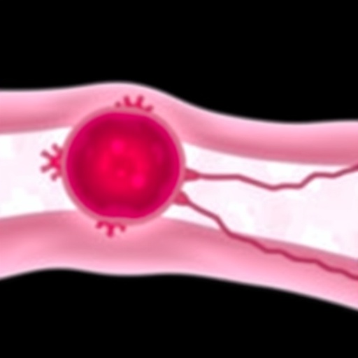
Image Credits: Shinichiro Sawa / Kyushu University
Keywords: bone marrow development, septoclasts, bone marrow stromal cells, RANKL, osteoclasts, fetal bone development, cartilage degradation, bone remodeling, LepR+ BMSCs, bone repair, cytokine signaling, skeletal regeneration
