


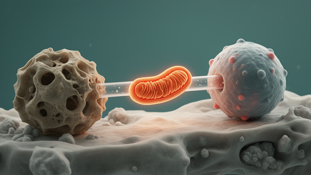
A 2026 review concludes that mitochondria move bidirectionally between skeletal and immune cells, functioning as context-dependent signals that can either support tissue homeostasis or amplify inflammation and bone damage. The authors distinguish this process from the separate intercellular transfer of mitochondrial components such as mitochondrial DNA, proteins or fragments.
The review, published in Frontiers in Endocrinology, synthesizes evidence on how skeletal cells—including bone marrow mesenchymal stem cells, osteoblasts and periodontal ligament stem cells—communicate with macrophages, osteoclast precursors, lymphocytes and other immune populations. It describes osteoimmunity as a cellular, molecular and metabolic network in which immune cells influence matrix remodeling while skeletal cells regulate immune responses.
Three routes were identified for transferring whole mitochondria between cells. Direct contact can involve actin-rich tunneling nanotubes, gap junctions or cell fusion, although the review notes debate over whether intact mitochondria pass directly through gap-junction channels. Extracellular vesicles can enclose intact mitochondria, fragments or mitochondrial DNA, while free mitochondria can be released into the extracellular space and captured by recipients through macropinocytosis or other phagocytic mechanisms.
Examples summarized in the review show that either direction of transfer can have different consequences. Osteolineage cells transferred mitochondria to myeloid osteoclast precursors were associated with improved glutathione metabolism, reduced ferroptosis and less bone resorption. Bone marrow mesenchymal stem cells also transferred mitochondria to T helper 17 cells, reducing interleukin-17 production and promoting conversion toward a regulatory T-cell phenotype. In contrast, mechanically compressed periodontal ligament fibroblasts released mitochondrial cargo in extracellular vesicles that activated the NLRP3 inflammasome in macrophages and induced caspase-1-dependent pyroptosis during orthodontic tooth movement.
Immune cells can also influence skeletal cells. Inflammatory macrophages transferred extracellular-vesicle-associated mitochondria to bone marrow mesenchymal stem cells, impairing osteogenic differentiation and exacerbating alveolar bone loss. By contrast, M2-like macrophages transferred functional mitochondria to injured periodontal ligament stem cells, activating p38 mitogen-activated protein kinase signaling and improving cell survival under inflammatory conditions. Separately, mechanical force activated Drp1 in macrophages and promoted EV-mediated mitochondrial transfer to mesenchymal stem cells through the CD200/CD200R axis, enhancing osteogenic differentiation in the cited experimental work.
The review emphasizes that the direction of transfer and the identity of the donor cell do not by themselves determine the outcome. Reactive oxygen species, hypoxia, inflammatory mediators and disrupted calcium homeostasis can alter mitochondrial dynamics and intercellular communication. The quality of donor mitochondria—including membrane potential, oxidative damage, cristae integrity and respiratory capacity—also matters. Functionally competent cargo can support oxidative phosphorylation, ATP production and redox balance, whereas damaged mitochondria or mitochondrial components can activate innate immune pathways. Recipient metabolic and lysosomal capacity and the surrounding microenvironment further shape the response.
The authors discuss pathological roles in osteoarthritis, osteoporosis and periodontitis. In osteoarthritis, mitochondrial material from stressed chondrocytes may be taken up by synovial macrophages and associated with TLR9, cGAS-STING and NLRP3 pathway activation, while M1-like macrophage EV-mitochondria can increase oxidative stress and chondrocyte dysfunction. In osteoporosis, impaired RIPK4-related mitochondrial dynamics and age-associated donor-cell dysfunction may weaken protective transfer to myeloid and osteogenic cells. In periodontitis, dysfunctional macrophage EV-mitochondria can suppress mesenchymal stem-cell osteogenic differentiation through the LCN2/OMA1/OPA1 pathway.
Four therapeutic categories are proposed: enhancing endogenous protective transfer, blocking pathological propagation, delivering exogenous mitochondria and engineering mitochondrial delivery. Reviewed interventions include functionalized iron oxide nanoparticles that improved aged macrophages in osteoporotic models, inhibitors of vesicle release or inflammasome signaling, and delivery of isolated mitochondria from mesenchymal stem cells to osteoarthritic chondrocytes. Engineered systems include hydrogels, responsive peptide complexes and mitochondrial carriers. These approaches are based mainly on cell-culture and animal evidence, and the review does not establish clinical efficacy in people.
Major limitations remain. Most evidence comes from in vitro studies and animal models, while standardized protocols are lacking for mitochondrial sourcing, isolation, dosing, characterization and safety evaluation. The review proposes a four-level evidence framework: direct in vivo tracing, direct in vitro imaging with functional testing, component-based evidence, and indirect evidence such as colocalization. Controls are needed to exclude dye leakage, cell fusion and uptake of apoptotic bodies, because fluorescence, mitochondrial DNA or mitochondrial proteins alone cannot prove transfer of intact mitochondria. The authors also call for assessment of purity, membrane integrity, respiratory activity, sterility, stability, biodistribution, immunogenicity and long-term toxicity before therapeutic mitochondria could advance toward clinical translation.
Subject of Research: Bidirectional mitochondrial transfer as a regulator of osteoimmune communication and bone disease
Article Title: Bidirectional mitochondrial transfer in the osteoimmune microenvironment: mechanisms and therapeutic perspectives
Article References: Huang, L., Chen, X., Guo, Y., Guo, F., Zhang, Y., Li, X., Dong, Z., & Kou, X. (2026). Bidirectional mitochondrial transfer in the osteoimmune microenvironment: mechanisms and therapeutic perspectives. Frontiers in Endocrinology, 17, Article 1978936. https://doi.org/10.3389/fendo.2026.1978936
Image Credits: AI Generated
DOI: 10.3389/fendo.2026.1978936
Keywords: mitochondrial transfer, osteoimmunity, bone remodeling, immunometabolism, mitochondrial quality control, extracellular vesicles, macrophages, mesenchymal stem cells, osteoporosis, osteoarthritis, periodontitis, Bidirectional
Cite Scienmag News
Drew Townsend. (October 21, 2026). Review maps bidirectional mitochondrial transfer across the osteoimmune microenvironment. Scienmag. https://scienmag.com/review-maps-bidirectional-mitochondrial-transfer-across-the-osteoimmune-microenvironment/
Drew Townsend. "Review maps bidirectional mitochondrial transfer across the osteoimmune microenvironment." Scienmag, 21 October 2026, https://scienmag.com/review-maps-bidirectional-mitochondrial-transfer-across-the-osteoimmune-microenvironment/. Accessed 21 October 2026.
Drew Townsend. "Review maps bidirectional mitochondrial transfer across the osteoimmune microenvironment." Scienmag. October 21, 2026. https://scienmag.com/review-maps-bidirectional-mitochondrial-transfer-across-the-osteoimmune-microenvironment/


