


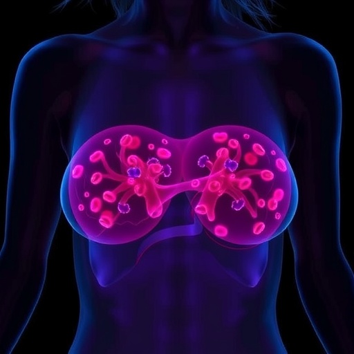
Breast cancer remains the most frequently diagnosed malignancy among women worldwide and is responsible for more than 650,000 deaths each year. Although modern oncology increasingly matches therapies to the molecular features of individual tumors, treatment success remains uneven. Patients with the same clinical subtype can experience dramatically different outcomes, suggesting that cancer cells alone do not determine how a disease progresses. The surrounding tumor microenvironment—a complex ecosystem of immune cells, fibroblasts, blood vessels and extracellular matrix—may be just as important. New research now points to mast cells, an immune population often associated with allergy and inflammation, as a potentially valuable source of prognostic information in a major breast cancer subtype.
The study, published in Genes & Immunity, examined transcriptional patterns linked to immune-cell infiltration in three publicly available breast cancer datasets. Rather than relying only on microscopic estimates of immune-cell abundance, the researchers analyzed gene-expression signatures that can indicate which immune populations are present in a tumor and whether those cells appear functionally activated. This approach, often called transcriptomic deconvolution, uses characteristic sets of genes to estimate the relative contribution of different cell types within a mixed tissue sample. The analysis revealed a consistent association between mast-cell states and clinical outcomes among patients with hormone receptor-positive, HER2-negative breast cancer.
Hormone receptor-positive, HER2-negative disease represents a large proportion of breast cancer cases. These tumors generally depend on estrogen or progesterone signaling for growth but lack overexpression of the HER2 protein, which can drive aggressive tumor behavior and can be targeted with specific drugs. Endocrine therapies are central to treatment, yet resistance and relapse remain significant clinical challenges. Unlike triple-negative and HER2-positive breast cancers, where higher levels of tumor-infiltrating lymphocytes often correlate with better survival, lymphocyte abundance has not shown the same predictive value in hormone receptor-positive, HER2-negative tumors. The new findings suggest that the immune biology of these cancers may need to be assessed through a wider lens.
The most striking observation involved the distinction between resting and activated mast cells. Greater infiltration by resting mast cells was repeatedly associated with improved survival indicators, whereas activated mast-cell signatures did not show the same favorable relationship. Mast cells are long-lived immune cells that reside in tissues and can release a broad range of biologically active substances, including histamine, proteases, cytokines and growth factors. Depending on their surroundings, these mediators can influence blood-vessel formation, tissue remodeling, inflammation and interactions between immune cells and cancer cells. Their effects are therefore highly context-dependent and cannot be classified as uniformly protective or harmful.
In the analyzed tumors, the presence of resting mast cells was inversely related to infiltration by other immune cells and to gene-expression markers associated with cancer-cell proliferation. At the same time, it correlated positively with stromal richness, meaning a greater contribution from the non-malignant structural compartment of the tumor. The stroma includes fibroblasts, connective-tissue proteins, small blood vessels and signaling molecules that provide both physical support and biochemical instructions to nearby cells. These relationships suggest that resting mast cells may be markers of a more organized or less aggressively inflamed tumor environment rather than direct agents of tumor destruction.
The finding is important because it shifts attention away from a simple question—how many immune cells are inside a tumor?—toward a more precise one: which immune cells are present, what state are they in, and how are they communicating with neighboring tissues? A tumor with abundant immune infiltration is not necessarily biologically favorable if those cells are suppressed, misdirected or associated with chronic inflammation. Conversely, a tumor with fewer conventional lymphocytes may still contain cellular networks that influence disease behavior through stromal organization and tissue repair pathways. Mast-cell activity could therefore complement established biomarkers rather than replace them.
One possible explanation is that interactions between resting mast cells and fibroblasts help shape a tumor microenvironment that is less supportive of rapid cancer-cell expansion. Fibroblasts can produce extracellular-matrix components and signaling factors that affect tumor stiffness, drug penetration, cell migration and immune access. Mast cells can influence fibroblast behavior through soluble mediators and direct cellular interactions. The balance between these populations may determine whether the stroma acts as a barrier, a scaffold for invasion or a relatively stable tissue compartment. However, the current study did not directly demonstrate such a mechanism. The proposed connection remains a biologically plausible hypothesis that will require laboratory and clinical investigation.
The results also carry potential implications for treatment sensitivity. Endocrine therapy, chemotherapy and emerging immune-based strategies can be affected by the physical and molecular properties of the tumor microenvironment. Dense or altered stroma may limit drug distribution, while inflammatory signaling can either stimulate immune attack or promote resistance. If mast-cell transcriptional states reliably identify tumors with distinct stromal and proliferative features, they could eventually contribute to risk stratification or help define groups for prospective clinical trials. Developing such applications would require standardized assays, validation in independent patient cohorts and proof that the signatures provide information beyond established clinical and genomic predictors.
The investigators emphasize that their conclusions are based on retrospective analyses of existing transcriptomic data. Gene-expression signatures estimate cellular abundance and functional state, but they do not provide the same direct evidence as tissue imaging, functional experiments or prospective treatment studies. An association between resting mast cells and longer survival does not prove that these cells cause better outcomes; they may instead be indicators of another protective feature of the tumor microenvironment. Even so, the consistency of the observation across three datasets strengthens the case for further research. By highlighting mast-cell state and stromal biology in hormone receptor-positive, HER2-negative breast cancer, the study opens a new avenue for understanding why apparently similar tumors can behave so differently—and why the next generation of personalized cancer care may need to profile not only malignant cells, but the entire ecosystem in which they survive.
Cite Scienmag News
Nathaniel Bowman. (July 31, 2026). Resting mast cell signature linked to improved outcomes in HR+HER2- breast cancer. Scienmag. https://scienmag.com/resting-mast-cell-signature-linked-to-improved-outcomes-in-hrher2-breast-cancer/
Nathaniel Bowman. "Resting mast cell signature linked to improved outcomes in HR+HER2- breast cancer." Scienmag, 31 July 2026, https://scienmag.com/resting-mast-cell-signature-linked-to-improved-outcomes-in-hrher2-breast-cancer/. Accessed 3 September 2026.
Nathaniel Bowman. "Resting mast cell signature linked to improved outcomes in HR+HER2- breast cancer." Scienmag. July 31, 2026. https://scienmag.com/resting-mast-cell-signature-linked-to-improved-outcomes-in-hrher2-breast-cancer/


