Endometriosis affects roughly 10 percent of women of reproductive age worldwide, an estimated 190 million people, yet its treatment remains stubbornly blunt. Hormonal drugs can ease symptoms but carry adverse events and high recurrence rates after discontinuation, while surgery can remove lesions at the cost of possible ovarian damage, with recurrence still looming. A team reporting in Materials Today Bio has now built a platinum-based nanomaterial designed to attack the disease from a different angle: not by suppressing estrogen, but by remodeling the oxidative and inflammatory environment that allows ectopic lesions to survive, invade, and fibrose.
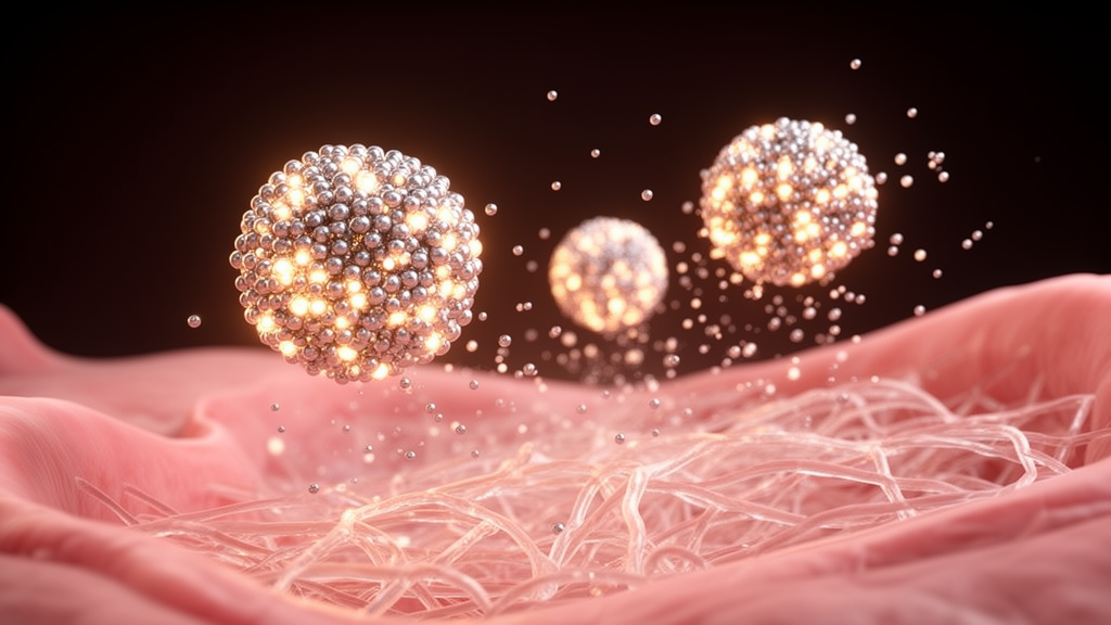
The material, called PtQDC, is a cluster of platinum quantum dots synthesized by a one-step hydrothermal method. Transmission electron microscopy showed nanoclusters 50 to 100 nanometers in diameter, composed of smaller quantum dots with a lattice spacing of 0.22 nanometers corresponding to the platinum (111) crystal plane. X-ray diffraction matched pure platinum, and X-ray photoelectron spectroscopy confirmed that the platinum was primarily in the zero-valent state. The design principle is bio-dissociability: rather than persisting as an intact particle, the cluster is meant to gradually come apart inside the lesion, releasing small quantum dot clusters that keep working within the diseased tissue.
To test that behavior, the researchers prepared fluid from ectopic lesions of model mice and suspended PtQDC in it. Over seven days, the clusters visibly thinned and lost edge integrity, shedding free platinum quantum dots of 2 to 4 nanometers. The zeta potential of the retained material shifted from −10.9 ± 1.6 millivolts initially to −20.2 ± 0.5 millivolts by day 7, consistent with the growing surface area produced by dissociation. The released quantum dots showed blue-green fluorescence under ultraviolet light, with peak emission at 465 nanometers under 370-nanometer excitation. The authors are careful to note that this dissociation does not establish biodegradation or systemic clearance; the fate, excretion pathways, and long-term safety of the released dots remain undetermined.
Functionally, PtQDC behaved as a broad-spectrum radical scavenger. In solution assays it removed hydrogen peroxide and the nitrogen-centered DPPH radical in concentration- and time-dependent fashion, and repeated cycling showed the activity held steady over four rounds. Electron spin resonance spectroscopy confirmed scavenging of PTIO radicals, singlet oxygen, superoxide anion, and hydroxyl radicals. The material also remained stable in phosphate-buffered saline, culture medium, and lesion tissue fluid, which the authors cite as evidence of good stability and biocompatibility across the tested environments.
The therapeutic concept hinges on a counterintuitive move: pushing diseased cells past their oxidative comfort zone into reductive stress. In 12Z endometriotic epithelial cells, PtQDC shifted the cells’ redox state in a concentration-dependent way, activating the GPX4–glutathione antioxidant system, raising the GSH/GSSG ratio, and nearly eliminating the oxidized lipid signal at the highest dose. But the reductive overshoot proved damaging. ATP levels fell, the mitochondrial membrane potential collapsed as JC-1 staining shifted from aggregates to monomers, and the cells lost their elongated, actin-organized architecture, rounding up as concentration increased. Cell viability stayed near control levels at 25 micrograms per milliliter but dropped significantly at 50 to 200 micrograms per milliliter.
That metabolic and mitochondrial dysfunction translated into functional impairment and cell death. Wound-healing assays showed progressively impaired migration at the higher doses, and cleaved caspase-3 fluorescence rose across the concentration series, indicating activation of apoptosis. Flow cytometry quantified the effect: the Annexin V-positive population climbed from about 7.1 percent in controls to 47.3 percent and 73.7 percent after treatment with 100 and 200 micrograms per milliliter, respectively, with a marked accumulation of late apoptotic cells.
In vivo, the team used a mouse model in which endometrial tissue from donor mice was transplanted into recipients, then treated the animals with PBS, a low dose, or a high dose of PtQDC. Treated mice showed significant decreases in both the number and volume of intra-abdominal lesions and reduced uterine adhesion scores compared with untreated model animals. In lesion tissue, malondialdehyde levels fell while superoxide dismutase activity rose, indicating alleviated oxidative stress. The authors report the treatment approach demonstrated effective biological safety during the therapy period, though the observation window was limited and longer-term safety was not established.
The inflammatory and vascular picture also shifted. In abdominal lavage fluid, PtQDC treatment, particularly at the high dose, reduced IL-6, IL-1β, and the pain-associated enzyme KLK1 while increasing the anti-inflammatory cytokine IL-10, with TNF-α unchanged. Immunofluorescence in lesion tissue confirmed dose-dependent decreases in proinflammatory factors and increased IL-10, which the authors interpret as immune rebalancing rather than simple suppression. Signals of pathological angiogenesis and hypoxia also fell: VEGF, the endothelial marker CD31, and HIF-1α all declined after treatment. Macrophage marker profiles changed too, with both CD86 and CD206 increasing and the CD86 rise more pronounced at the high dose, a shift the authors suggest may favor an M1-associated phenotype that restricts lesion progression. KLK1 and substance P, mediators of nociceptive sensitization, were both reduced in lesions, hinting at attenuated pain signaling, though no behavioral pain outcomes were reported.
Transcriptomic analysis added a molecular layer. RNA sequencing of treated versus model tissue identified 108 differentially expressed genes, and network analysis distilled 41 core genes, with Ptgs2, Postn, and Mmp3 showing the highest degree values. These three encode, respectively, a prostaglandin-synthesizing driver of inflammation and pain, an extracellular matrix protein promoting fibrosis and adhesion, and a matrix metalloproteinase that helps ectopic cells invade. In a publicly available dataset of 408 clinical samples, all three genes were elevated in endometriotic foci, especially deep infiltrating and ovarian lesions, and PtQDC downregulated all three in mouse lesions. Among 74 patients staged by disease severity, only ALDH1A3 was significantly elevated in severe cases, and PtQDC also downregulated it, pointing toward a possible role for retinol and vitamin A metabolism that the authors flag as a new research direction.
The authors position PtQDC as a multitarget, nonhormonal strategy whose sequential actions—radical scavenging, inflammatory rebalancing, and inhibition of angiogenesis and fibrosis—address the microenvironment that sustains endometriotic lesions. The evidence so far is preclinical: cell lines and a mouse transplant model with small group sizes, no human dosing, and unresolved questions about the long-term fate of released platinum quantum dots and lesion recurrence after treatment withdrawal. The team itself notes that durability of benefit and recurrence following treatment cessation need future study, and that limited sequencing sample size may have masked steroid-pathway effects. Still, by demonstrating that a self-dismantling nanozyme can simultaneously blunt oxidative stress, inflammation, neovascularization, and invasion-associated gene expression in model lesions, the study opens a concrete path toward nonhormonal endometriosis therapy—one that now must survive the longer, harder tests of preclinical safety and translational evaluation.
Subject of Research: A bio-dissociable platinum nanozyme that scavenges reactive species to treat endometriosis in cell and mouse models
Article Title: Bio-dissociable platinum quantum dot cluster nanozymes for nonhormonal endometriosis therapy through microenvironment remodeling
Article References: Cao, C., Ding, J., Hao, H., Jiao, Y., Cao, Y., Fu, Y., Cui, X., Gao, D., Gao, Y., Wang, Y., Yu, C., Wang, T., & Liu, Y. (2026). Bio-dissociable platinum quantum dot cluster nanozymes for nonhormonal endometriosis therapy through microenvironment remodeling. Materials Today Bio, 41, Article 103785. https://doi.org/10.1016/j.mtbio.2026.103785
Image Credits: AI Generated
DOI: 10.1016/j.mtbio.2026.103785
Keywords: endometriosis, nanozymes, platinum quantum dots, reactive oxygen species, oxidative stress, inflammation, angiogenesis, mouse model, nonhormonal therapy, microenvironment remodeling, transcriptomics, preclinical research
Cite Scienmag News
Katie Riggs. (October 28, 2026). Research report: Bio-dissociable platinum quantum dot cluster nanozymes for nonhormonal endometriosis therapy through microenvironment remodeling. Scienmag. https://scienmag.com/research-report-bio-dissociable-platinum-quantum-dot-cluster-nanozymes-for-nonhormonal-endometriosis-therapy-through-microenvironment-remodeling/
Katie Riggs. "Research report: Bio-dissociable platinum quantum dot cluster nanozymes for nonhormonal endometriosis therapy through microenvironment remodeling." Scienmag, 28 October 2026, https://scienmag.com/research-report-bio-dissociable-platinum-quantum-dot-cluster-nanozymes-for-nonhormonal-endometriosis-therapy-through-microenvironment-remodeling/. Accessed 28 October 2026.
Katie Riggs. "Research report: Bio-dissociable platinum quantum dot cluster nanozymes for nonhormonal endometriosis therapy through microenvironment remodeling." Scienmag. October 28, 2026. https://scienmag.com/research-report-bio-dissociable-platinum-quantum-dot-cluster-nanozymes-for-nonhormonal-endometriosis-therapy-through-microenvironment-remodeling/