


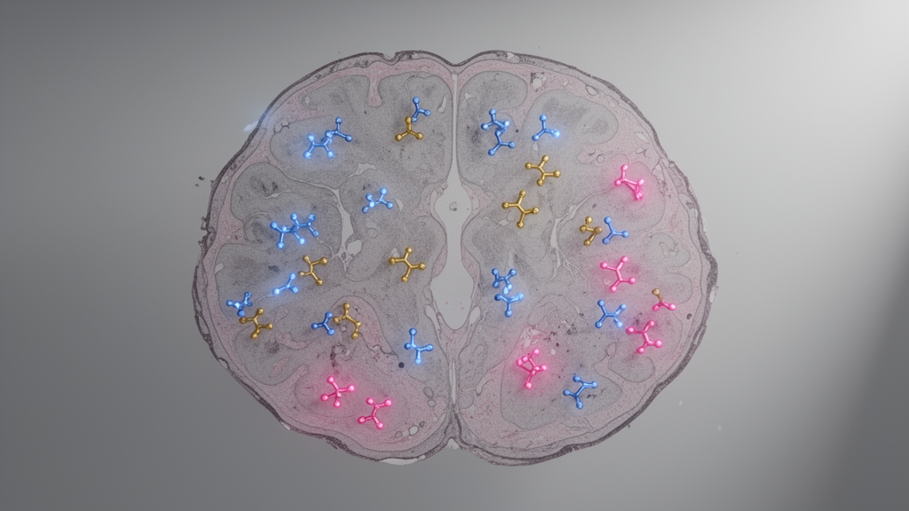
A University at Buffalo research team has created thousands of protein “satellite maps” that show where monoclonal antibodies and more than 6,000 brain proteins are distributed across entire mouse brain tissue sections. The method, called hexagonal micro-scaffold assisted spatial proteomics, or hex-MASP, combines a three-dimensionally printed scaffold with ultrasensitive mass spectrometry to preserve and reconstruct the location of proteins throughout treated tissue. Demonstrated in a peer-reviewed animal study published in the Proceedings of the National Academy of Sciences, the work provides a detailed view of antibody distribution alongside the molecular landscape of the brain.
The researchers said the maps could help identify therapeutic targets, reveal patterns associated with disease or treatment, and guide the development of medicines, but the experiments did not test benefits in people. Monoclonal antibodies are medicines designed to bind specific protein targets, and their uneven distribution within an organ can be associated with uneven responses across tissue. In the experiment, the researchers used the platform to determine where administered antibodies appeared and where more than 6,000 brain proteins were located.
Each protein had its own distribution map, while the antibody maps provided a parallel view of the treatment within the same tissue sections. Viewed together, the maps connected the location of a medicine with the locations of potential targets and the surrounding tissue response. At the heart of the method is a scaffold containing tiny hexagonal wells into which a tissue section is pressed.
The wells divide the section into numerous microsamples while retaining the original position of each sample. This spatial record is essential because it allows later molecular measurements to be returned to the part of the tissue from which they came. Earlier versions of the technique used square compartments, which the team said could bend, break or collapse when pressed into tougher tissues such as the heart or kidney.
The researchers selected a hexagonal architecture because it provides a stronger structure and more reliably preserves the tissue’s spatial arrangement during partitioning. After the scaffold separates a section into position-retaining microsamples, ultrasensitive mass spectrometry measures proteins in those individual samples. The resulting measurements are reconstructed into maps showing the distribution of each detected protein across the full tissue section.
In the mouse-brain application, this workflow generated maps for the therapeutic antibodies and for more than 6,000 other brain proteins. The authors describe each protein map as one part of the tissue’s biological story, with the strongest information emerging when thousands of maps are examined together. The motivation for the technology comes from the uneven way medicines can reach different regions of an organ.
Qu said that when a drug enters a tissue, it generally does not do so uniformly, creating variations in biological response. Hex-MASP was designed to make that spatial information available. By mapping an antibody alongside thousands of proteins, researchers can examine whether the treatment reaches tissue regions where its targets are present and then study how those regions respond.
The reported findings establish the mapping capability in mouse brain tissue; they do not demonstrate that the method can determine the causes of differences in treatment benefit. Alzheimer’s disease provides one proposed area of application. Qu said researchers have spent decades developing treatments intended to clear amyloid plaques, with the hope that doing so would substantially improve patients’ outcomes.
One pharmaceutical company developed a series of antibodies that cleared most plaque, but their clinical trials produced a cognitive benefit that did not match the extent of plaque removal, according to Qu. Antibodies that have since received approval only moderately slow cognitive decline rather than reverse it. Qu said this pattern appears to show that targeting amyloid alone does not address enough of the biology driving disease progression, although the supplied study did not directly test the clinical effects of the new mapping method.
The proposed next step is to map the abundance and modification status of thousands of proteins across brain tissue at different stages of Alzheimer’s disease. Such maps could reveal disease processes that researchers have not previously considered and help identify changes that are most important or offer better opportunities for treatment. The authors also propose using hex-MASP to evaluate medicines developed against those newly identified targets.
By mapping a drug candidate together with the brain’s protein response, researchers could check whether a candidate changes the biology it was intended to change. This use remains a development objective rather than a clinical outcome demonstrated by the mouse-brain experiment, and the platform has not been shown to predict responses in individual patients or to improve treatment selection in human medicine. Qu’s laboratory has already identified candidates it describes as promising for Alzheimer’s drug targets, with further research underway.
That finding is a statement about the laboratory’s ongoing target-discovery work, not evidence that the candidates are effective treatments. The published study did not report testing such candidates for safety or efficacy in people, and it did not establish that any mapped protein causes treatment benefit. Instead, the demonstrated result is a preclinical measurement technology capable of producing tissue-wide protein and antibody maps from mouse brain sections.
Further development is needed to determine how reliably the method performs across other tissues, diseases and classes of medicines, as well as how its findings translate into decisions about drug design and delivery. The team plans to extend hex-MASP to additional diseases and types of medicines. The group was led by Jun Qu, a professor of pharmaceutical sciences in the University at Buffalo School of Pharmacy and Pharmaceutical Sciences, and included colleagues in pharmaceutical sciences and the Department of Industrial and Systems Engineering.
The work was supported by National Institutes of Health grant U01 DK137113 and a Center for Protein Therapeutics consortium grant, with data obtained through the University at Buffalo Center for Proteomics and Bioanalysis. Their mouse-brain results provide the experimental foundation for that goal, while the potential to improve therapies for patients remains to be tested.
Subject of Research: Spatial proteomic mapping of monoclonal antibody and brain-protein distribution in treated mice
Article Title: Creating ‘satellite maps’ of proteins could transform drug therapies
Article References: Creating ‘satellite maps’ of proteins could transform drug therapies. Original publication (Bibliographic details unavailable.)
Image Credits: AI Generated
DOI: Not provided
Keywords: spatial proteomics, hex-MASP, mouse brain, monoclonal antibodies, protein mapping, mass spectrometry, tissue distribution, drug development, Alzheimer’s disease, Creating, satellite, maps
Cite Scienmag News
Cassandra Pierce. (October 26, 2026). Protein Maps Chart Antibodies Across Mouse Brain Tissue. Scienmag. https://scienmag.com/protein-maps-chart-antibodies-across-mouse-brain-tissue/
Cassandra Pierce. "Protein Maps Chart Antibodies Across Mouse Brain Tissue." Scienmag, 26 October 2026, https://scienmag.com/protein-maps-chart-antibodies-across-mouse-brain-tissue/. Accessed 26 October 2026.
Cassandra Pierce. "Protein Maps Chart Antibodies Across Mouse Brain Tissue." Scienmag. October 26, 2026. https://scienmag.com/protein-maps-chart-antibodies-across-mouse-brain-tissue/


