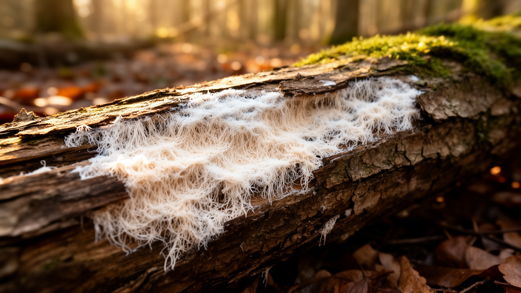
When the white rot fungus Phlebia radiata is hit with a sudden dose of hydrogen peroxide while growing on birch wood, its genome does not simply shut down. Instead, researchers report in Fungal Biology and Biotechnology that the fungus mounts a highly specific transcriptional response, one that differs markedly depending on whether the cultures are kept in ordinary air or in a low-oxygen atmosphere that mimics the interior of decaying wood.
The team, based at the University of Helsinki, cultivated the fungus on silver birch sapwood sawdust in static laboratory flasks. On the tenth day of cultivation, half of the cultures received hydrogen peroxide at a final concentration of 0.1 percent by weight, equivalent to 35 millimolar, while the rest received only sterile water. Half of the flasks had been sealed with tight rubber plugs fitted with inlet valves five days earlier, dropping the headspace oxygen to microaerophilic levels, while the remaining cultures kept ventilating cellulose stoppers and were assumed to stay near atmospheric 21 percent oxygen. The researchers sampled cultures before treatment, 18 hours afterwards, and again four days later, with five biological replicate flasks per condition and timepoint.
RNA sequencing of 24 samples mapped reads to 12,450 of the 14,067 annotated genes in the updated P. radiata genome assembly. Combining both atmospheres, 762 genes were significantly differentially expressed 18 hours after the peroxide shock, using a false discovery rate threshold of 0.05 and an absolute log2 fold change threshold of 1. The response split along atmospheric lines: in the aerobic cultures, 267 genes were upregulated and 236 downregulated, with 153 upregulated genes unique to that condition, whereas the low-oxygen cultures showed 348 downregulated genes, 185 of them unique to that condition, and a third of those downregulated genes encoded proteins of unknown function.
Only 113 genes were upregulated and 162 downregulated under both atmospheres, and just one gene moved in opposite directions. Among the shared upregulated genes, small secreted proteins dominated. These are proteins shorter than 250 amino acids carrying an N-terminal secretion signal and, in most cases, no recognizable protein domain. Twenty-four of the 113 shared upregulated genes encoded such proteins, and many ranked among the fifty most drastically induced genes. Six of them encode tiny 68-amino-acid proteins that share 76.4 to 100 percent amino-acid identity with one another. After the SSPs came short-chain dehydrogenase/reductases, aldo-keto reductases, GNAT family acetyltransferases, and carbohydrate-active enzymes of the CAZy classification.
The redox enzyme response was particularly striking. Seven of the 59 predicted aldo-keto reductase genes were upregulated under both conditions, spanning the AKR1-5-like, AKR7, AKR8, AKR9 and AKR13 subfamilies, and several remained highly expressed four days after the shock. None carried a secretion signal, indicating intracellular function. The authors suggest these enzymes, along with retinol dehydrogenase-like short-chain dehydrogenase/reductases acting on antioxidant molecules such as ubiquinone and retinol, may reflect remediation of lipid peroxidation caused by hydrogen peroxide and other reactive oxygen species. An Old Yellow Enzyme gene, a protein family linked to reactive oxygen species resistance, also appeared among the most upregulated genes.
Carbon metabolism told the opposite story. Under both atmospheres, the oxidative treatment suppressed the pentose catabolic pathway, glycerol metabolism, and the routes leading to ethanol and acetate. Downregulated genes included a xylulokinase, a xylulose 5-phosphate phosphoketolase, the sole gene of its kind in the genome, several glycerol dehydrogenases, a dihydroxyacetone kinase, a glycogen phosphorylase, a phosphoglycerate kinase, a pyruvate decarboxylase and an acetate kinase. Gene set enrichment analysis across all KEGG-mapped genes confirmed that oxidative phosphorylation, pentose and glucuronate interconversions, fructose and mannose metabolism, and protein processing in the endoplasmic reticulum were generally downregulated under both conditions, pointing to a broad suppression of primary metabolism as an immediate shock response.
The atmosphere shaped which side of the metabolism took the hit. In aerobic cultures, enrichment analysis flagged glycolysis and gluconeogenesis, pyruvate metabolism, steroid biosynthesis, carbon metabolism, oxidative phosphorylation, propanoate metabolism, and fructose and mannose metabolism as downregulated, while proteasome and lysine biosynthesis genes trended upward. In the low-oxygen cultures, the effect reached deeper into essential cell functions and repair mechanisms. The low-oxygen flasks were not anaerobic: headspace oxygen fell from about 9 percent on day 10 to 4 percent by day 14, with a slight rise to 10 percent after peroxide addition, while carbon dioxide accumulated to 12 to 14 percent, a signature of fermentative metabolism.
Some findings ran against expectations. Seven heat shock protein genes were among the shared downregulated genes, five of them encoding small Hsp20-family proteins, and a ClpB/HSP104 chaperone gene was also repressed, along with a heat shock factor family protein and a hybrid histidine kinase. The authors propose that the oxidative treatment may have inflicted less severe intracellular protein damage than heat stress would, so that costly protein-repair machinery was not needed. Meanwhile, peroxide wiped out extracellular enzyme activity: laccase and manganese peroxidase activities of roughly 17 and 1 nanokatals per milliliter on day 10 were undetectable 18 hours after treatment, recovering only to a tenth or a twentieth of their former levels by day 14. Xylanase activity, by contrast, rose transiently after treatment, especially in aerobic cultures, before both treated and untreated cultures returned to a similar range of about 4 to 5 nanokatals per milliliter.
Metabolite measurements reinforced the picture of a fungus that survived the shock. Veratryl alcohol, a well-characterized natural product of this species, continued to accumulate in cultures under both atmospheres regardless of treatment, which the authors read as evidence of fungal vitality. Veratraldehyde concentrations spiked 18 hours after peroxide addition but returned to baseline by day 14, whereas untreated cultures showed steady accumulation. DPPH antioxidant activity in culture extracts increased slightly after treatment under both conditions, and iron reduction capacity, which was far lower in fungal cultures than in no-fungus wood substrate controls, dropped sharply in the controls after peroxide addition, indicating that the treatment eliminated wood and medium-derived chemical reductants.
The study’s conclusion is that P. radiata does not respond to oxidative shock with wholesale transcriptomic silencing but with a targeted program: induction of small secreted proteins, aldo-keto reductases and a few hydrolytic enzymes, alongside repression of pentose and glycerol catabolism and sugar transporter genes, including four of the genome’s 25 hexose MFS transporters. The authors interpret the transporter and pathway downregulation as consistent with the shock releasing extra hexoses and saccharides from the wood substrate, reducing the need for uptake and pentose utilization. Because the two atmospheres produced different responses, the researchers attribute the divergence to contrasting metabolic states before the shock, visible in enzyme, metabolite and gas profiles. Four days after treatment, only 42 genes remained differentially expressed between treated and untreated cultures, and just three genes were differentially expressed at both timepoints under both conditions, indicating that the fungus largely recovered its baseline expression within days. The work leaves the functions of most of the responsive small secreted proteins unknown, but suggests they participate in rapid metabolic adaptation and reactive oxygen species mitigation in wood-decay fungi, a group whose stress biology remains far less studied than that of pathogenic species.
Subject of Research: Transcriptomic and metabolic responses of the white rot fungus Phlebia radiata to oxidative stress on wood substrate under different atmospheric conditions
Article Title: Oxidative stress impacts carbon metabolism and induces upregulation of redox enzymes andsmall secreted proteins in the wood-decay fungus
Article References: Österman-Udd, J., Kiviniemi, E. A., Simojoki, A., Wahlsten, M., & Lundell, T. (2026). Oxidative stress impacts carbon metabolism and induces upregulation of redox enzymes andsmall secreted proteins in the wood-decay fungus. Fungal Biology and Biotechnology, 13(1), Article 17. https://doi.org/10.1186/s40694-026-00222-2
Image Credits: AI Generated
DOI: 10.1186/s40694-026-00222-2
Keywords: Phlebia radiata, oxidative stress, hydrogen peroxide, wood decay fungi, RNA-Seq, small secreted proteins, aldo-keto reductases, carbon metabolism, hypoxia, lignocellulose, gene expression, white rot
Cite Scienmag News
Juliet Wilcox. (October 28, 2026). Oxidative shock rewires gene expression in white rot fungus on wood. Scienmag. https://scienmag.com/oxidative-shock-rewires-gene-expression-in-white-rot-fungus-on-wood/
Juliet Wilcox. "Oxidative shock rewires gene expression in white rot fungus on wood." Scienmag, 28 October 2026, https://scienmag.com/oxidative-shock-rewires-gene-expression-in-white-rot-fungus-on-wood/. Accessed 28 October 2026.
Juliet Wilcox. "Oxidative shock rewires gene expression in white rot fungus on wood." Scienmag. October 28, 2026. https://scienmag.com/oxidative-shock-rewires-gene-expression-in-white-rot-fungus-on-wood/