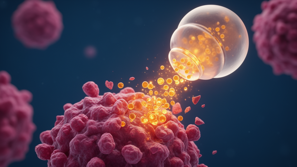
Researchers have engineered a temperature-sensitive nanoparticle that carries plumbagin, a plant-derived naphthoquinone with known anticancer activity, and releases it in a way that responds to the slight temperature differences between healthy tissue and the tumour microenvironment. In a study published in Biotechnology for Sustainable Materials, a team from India fabricated four plumbagin-loaded nanoformulations using chitosan and polylactic acid (PLA) as polymeric matrices, and identified one formulation, designated F3, as the most promising candidate. In laboratory tests with LPS-induced inflammation in SiHa cervical cancer cells, the mid dose of 0.75 micrograms per millilitre inhibited cell migration, induced apoptosis and autophagy, and arrested cells in the G2/M phase of the cell cycle. The work remains at the in vitro stage, and the authors themselves stress that animal studies and clinical investigations are needed before any therapeutic use.
The motivation behind the study lies in the limitations of current cervical cancer treatment. Cervical cancer is the fourth most prevalent cancer in women worldwide according to the WHO, and roughly 70 percent of cases worldwide are driven by infection with HPV-16 or HPV-18. The main treatment options, which include surgery, radiation therapy, chemotherapy and targeted or immunotherapy, can be effective but carry a significant risk of severe systemic side effects because of high and repeated doses, imposing an economic and physiological burden on patients. Plumbagin, a naphthoquinone compound derived from the Plumbaginaceae family, has shown anticancer activity against cervical cancer cells in several studies, but its poor solubility and low bioavailability have limited its practical use. Encapsulating the compound in a polymeric nanoparticle is one strategy to overcome those constraints.
The team prepared the four formulations using an ionotropic gelation technique, combining two parts chitosan with one part PLA as excipients and one part plumbagin as the active ingredient, with the PLA used in either crystalline or amorphous form. The physical characterisation of these particles, including their pH-responsive behaviour, antimicrobial activity and cytotoxic potential, had been reported in a previous publication; the new study focused on their thermoresponsive properties and anticancer bioactivity. The central design idea is that solid tumours run slightly warmer than surrounding healthy tissue because of increased metabolic heat generation and altered blood perfusion, so a carrier that changes structure across that narrow temperature window could, in principle, release its cargo preferentially at the diseased site.
To characterise that behaviour, the researchers measured the lower critical solution temperature (LCST) of each formulation by UV-visible spectrophotometry at 450 nanometres, heating samples from 10 to 60 degrees Celsius and tracking light transmittance. The phase transition of formulations F1, F3 and F4 occurred around 38 to 40 degrees Celsius, while F2 transitioned at 35 degrees Celsius. The nanoformulations were then exposed to two temperatures, 37 degrees Celsius representing normal body temperature and 39 degrees Celsius representing a diseased condition, and their drug release profiles were measured in simulated vaginal fluid using a dialysis bag technique over 24 hours.
Formulation F3 stood out clearly. It released 54.3 percent of its plumbagin payload at 37 degrees Celsius and 31.7 percent at 39 degrees Celsius, the highest cumulative release among the four formulations. By comparison, F1 released 32.4 and 28.06 percent, F2 released 21.9 and 17.5 percent, and F4 released 31.4 and 14.8 percent at the two temperatures respectively. Free, unencapsulated plumbagin showed essentially temperature-independent release, at 25.5 percent at 37 degrees and 26.2 percent at 39 degrees, confirming that the thermoresponsive behaviour came from the polymeric carrier rather than the drug itself. Interestingly, the formulation released more drug at the normal physiological temperature than at the elevated diseased-condition temperature, which the authors attribute to structural rearrangements of the thermoresponsive polymer matrix at temperatures approaching or exceeding the LCST.
Kinetic modelling showed that all four formulations followed the Higuchi model of release at both temperatures, indicating that the drug diffuses steadily out of the polymeric matrix rather than being released by matrix erosion alone. Korsmeyer–Peppas analysis yielded release exponent values of 0.40 at 37 degrees and 0.39 at 39 degrees, consistent with Fickian diffusion. Thermal analysis by TGA-DSC revealed a shift of the endothermic peak towards lower temperatures and a broader transition at 39 degrees, suggesting increased polymer chain mobility and a phase transition within the network. X-ray diffraction showed temperature-dependent changes in crystallinity, with F3 restoring the characteristic plumbagin diffraction peak at reduced intensity, and scanning electron microscopy revealed surface erosion and loss of compact morphology after thermal treatment, with more pronounced changes under the diseased condition.
With F3 selected on the basis of its release profile, the team turned to biological testing in SiHa cells, an HPV-16-positive cervical cancer cell line, in which inflammation was induced with lipopolysaccharide (LPS) to mimic inflammation-driven features of the tumour microenvironment. Three doses were tested: 0.25, 0.75 and 1.5 micrograms per millilitre, with paclitaxel and dexamethasone as reference standards. In a scratch assay, the mid and high doses abolished cell migration entirely, with cell death apparent at 12 and 24 hours, while the low dose allowed only minimal migration before cell death began at 24 hours. Paclitaxel followed a similar pattern, whereas the normal control, LPS-treated and dexamethasone-treated cells migrated readily.
Flow cytometric analysis with Annexin-V/PI staining showed that 64.5 percent of cells treated at the high dose and 60.9 percent at the mid dose were in a necrotic state, compared with 22 percent at the low dose; paclitaxel produced 10.8 percent necrotic cells. The authors suggest the necrotic component may reflect excessive cellular stress, generation of reactive oxygen species and impaired ATP production from uncoupled mitochondrial oxidation. Cell cycle analysis by propidium iodide staining revealed a dose-dependent accumulation of cells in the G2/M phase across all treated groups, along with a sub-G1 population indicative of cell death. Gene expression analysis supported these observations: the pro-apoptotic gene BAX was significantly upregulated and the anti-apoptotic gene BCL2 significantly downregulated at the mid dose, while CDK1 and Cyclin B1, key regulators of the G2/M transition, were downregulated in mid- and high-dose groups. Autophagy markers Beclin and LC3 were significantly increased at the low and mid doses, and MDC staining confirmed the formation of autophagic vacuoles.
Immunoblot analysis at the mid dose added a protein-level dimension to the picture. Total caspase-9 was downregulated in the nanoformulation-treated group, but its activated form, cleaved caspase-9, was significantly upregulated compared with the normal control, a pattern consistent with activation of the intrinsic apoptotic pathway. Paclitaxel showed no significant difference in cleaved caspase-9 in this assay. Taken together, the molecular and imaging data point to a formulation that simultaneously promotes apoptosis, induces autophagy and blocks cell-cycle progression in the treated cells, with the mid dose of 0.75 micrograms per millilitre consistently showing the strongest effect among the three doses tested.
The authors are careful to frame the work as proof of concept. They note that batch-to-batch consistency in parameters such as zeta potential, particle size, encapsulation efficiency and drug release will be essential for any scale-up, and that changes in process parameters during translation from laboratory protocol to commercial production could affect reproducibility. They also emphasise that validation in animal models and ultimately clinical investigations will be required to establish translational relevance. Within those limits, the study demonstrates that a chitosan–PLA thermoresponsive carrier can tune plumbagin release across a physiologically meaningful temperature range and deliver measurable anticancer effects in an in vitro model of inflamed cervical cancer cells, offering a starting point for further development of targeted vaginal delivery systems for cervical cancer treatment.
Subject of Research: Thermoresponsive plumbagin-loaded chitosan–PLA nanoformulations for cervical cancer drug delivery
Article Title: Physicochemical and biomolecular characterisation of plumbagin incorporated thermoresponsive nanoformulation
Article References: Vasanthi, H. R., Subi, M. T. M., Priyadharshini, S., & Rajesh, S. (2026). Physicochemical and biomolecular characterisation of plumbagin incorporated thermoresponsive nanoformulation. Biotechnology for Sustainable Materials, 3(1), Article 11. https://doi.org/10.1186/s44316-026-00062-5
Image Credits: AI Generated
DOI: 10.1186/s44316-026-00062-5
Keywords: plumbagin, thermoresponsive nanoformulation, chitosan, polylactic acid, cervical cancer, SiHa cells, drug release, Higuchi model, apoptosis, autophagy, G2/M arrest, LCST
Cite Scienmag News
Nathaniel Bowman. (October 28, 2026). Plumbagin-loaded thermoresponsive nanoparticles show anticancer effects in cervical cancer cells. Scienmag. https://scienmag.com/plumbagin-loaded-thermoresponsive-nanoparticles-show-anticancer-effects-in-cervical-cancer-cells/
Nathaniel Bowman. "Plumbagin-loaded thermoresponsive nanoparticles show anticancer effects in cervical cancer cells." Scienmag, 28 October 2026, https://scienmag.com/plumbagin-loaded-thermoresponsive-nanoparticles-show-anticancer-effects-in-cervical-cancer-cells/. Accessed 28 October 2026.
Nathaniel Bowman. "Plumbagin-loaded thermoresponsive nanoparticles show anticancer effects in cervical cancer cells." Scienmag. October 28, 2026. https://scienmag.com/plumbagin-loaded-thermoresponsive-nanoparticles-show-anticancer-effects-in-cervical-cancer-cells/