


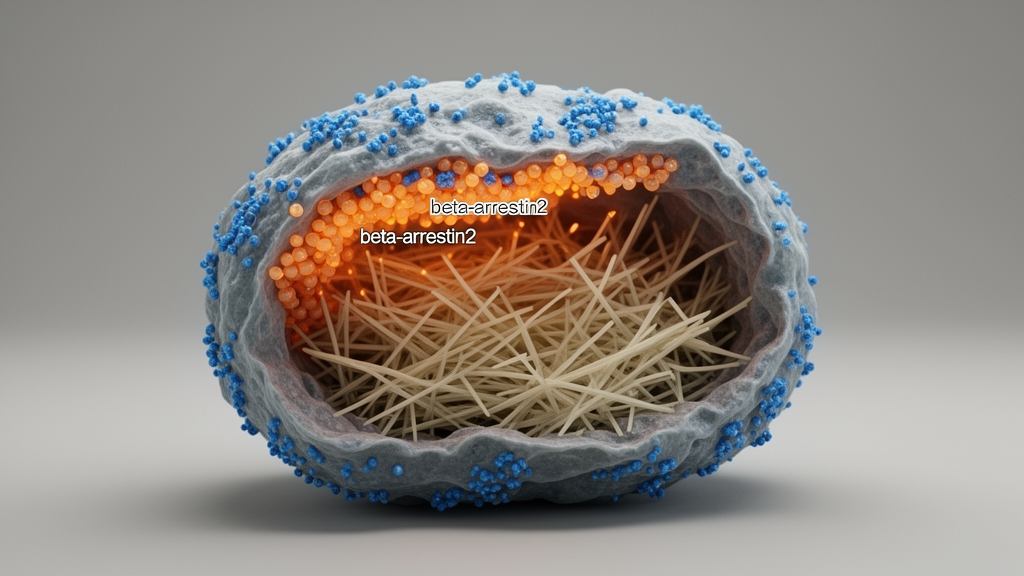
For decades, cardiologists have treated the failing heart as a single functional unit, directing therapies at the muscle tissue that pumps blood while paying far less attention to the cells that build and repair its structural scaffolding. A new study published in the Journal of Cellular and Molecular Medicine by Xu and colleagues now argues that this oversight may have real pharmacological consequences. The research demonstrates that the β3-adrenergic receptor, long celebrated as a cardioprotective target, can drive a profibrotic program in cardiac fibroblasts through a signaling route that bypasses the classical G-protein machinery entirely. Instead, the receptor recruits the adaptor protein β-arrestin2, which activates the JNK/c-Jun pathway and upregulates transforming growth factor β1, a master driver of collagen deposition and scar formation. The finding reframes the fibroblast not as a passive bystander in sympathetic overdrive but as an independent pharmacological compartment whose responses to the same hormone can differ radically from those of neighboring cardiomyocytes.
The β3-adrenergic receptor occupies an unusual position in cardiovascular pharmacology. Unlike its β1 and β2 cousins, which stimulate contractility and heart rate through stimulatory G proteins, the β3 subtype has been associated with inhibitory Gi coupling, activation of nitric oxide synthase, and negative inotropic effects that can shield an overworked myocardium from excessive catecholamine stress. Studies in genetically modified mice and in isolated cardiomyocytes have repeatedly linked enhanced β3 receptor expression to protection against hypertrophic remodeling, oxidant stress, and neurohormone-induced damage. This body of evidence fueled optimism that β3 agonists might one day complement or refine conventional β-blockade. The new work punctures that uniform narrative. In fibroblasts, the very same receptor behaves in an entirely different manner, triggering a cascade that promotes the transformation of quiescent fibroblasts into collagen-secreting myofibroblasts, the cellular engines of myocardial fibrosis.
The mechanistic core of the study lies in the concept of biased signaling, one of the most actively pursued ideas in contemporary G-protein-coupled receptor pharmacology. A single receptor can adopt multiple active conformations, each preferentially stabilizing a different transducer: a heterotrimeric G protein on one hand, or an arrestin scaffold on the other. Xu and colleagues show that in cardiac fibroblasts, β3-adrenergic receptor activation preferentially engages β-arrestin2, and that this recruitment is both necessary and sufficient to activate the c-Jun N-terminal kinase pathway. Activated c-Jun then drives transcriptional upregulation of TGF-β1, the cytokine that orchestrates fibrotic remodeling across virtually every organ system. Notably, the authors report that this profibrotic output appears to operate independently of classical Gi protein coupling, meaning the receptor’s two canonical outputs can be dissociated within a single cell type. That dissociation is precisely what makes the finding exciting for drug developers, because it suggests the theoretical possibility of ligands that preserve beneficial Gi signaling while silencing the arrestin arm.
The timing of the report is significant. The pharmaceutical industry has invested heavily in biased ligands, molecules engineered to steer receptors toward therapeutically favorable pathways, with angiotensin receptor agonists and opioid candidates among the most prominent examples. The new data supply a mechanistic rationale for extending this strategy to the adrenergic system in cardiac disease. If a β3-selective ligand could be designed to activate Gi-dependent nitric oxide signaling in cardiomyocytes while blocking β-arrestin2 recruitment in fibroblasts, it might in principle halt myocardial fibrosis without compromising contractility, a combination that blunt β-blockade cannot achieve. The study also helps reconcile a literature that has long appeared contradictory: some groups reported that β3 receptor deletion worsens ventricular remodeling, while others found that β3 agonism exacerbates atrial structural remodeling through nitric oxide uncoupling. Cell-specific signaling bias offers a coherent framework for understanding how both observations could be true at once.
Yet the study carries methodological caveats that temper its immediate translational promise, and these deserve careful scrutiny. The experimental system relies on receptor overexpression to stimulate β3-adrenergic signaling, an approach known to distort receptor biology in consequential ways. Artificially elevated receptor abundance can alter subcellular distribution, receptor stoichiometry, and the recruitment of adaptor proteins, producing non-physiological receptor pools that do not mirror the predominantly cell-surface localization of endogenous receptors. A recent study from the Lefkowitz laboratory demonstrated that biased signaling at a G-protein-coupled receptor can be lost entirely in overexpressed systems, raising the possibility that the profibrotic phenotype observed here reflects receptor abundance and clustering rather than an intrinsic property of the receptor’s signaling mechanism. Until the pathway is confirmed at endogenous expression levels, the claim of bona fide biased agonism remains an inference rather than a demonstrated fact.
A second limitation concerns the structural basis of the arrestin interaction. β-arrestin2 recruitment to G-protein-coupled receptors is governed by receptor phosphorylation, conformation, and cellular context, with specific serine and threonine residues serving as docking signals once phosphorylated by G-protein-coupled receptor kinases. The study does not map these phosphorylation sites on the β3-adrenoceptor, leaving open the question of which molecular features of the receptor determine its arrestin preference in fibroblasts. Without such a map, it is impossible to define the precise structural switch that controls the profibrotic transition, or to predict whether the same switch operates in other cell types and species. Future work should identify the responsible phosphorylation residues and consider how tissue-specific microenvironmental cues, including mechanical stress, hypoxia, and inflammatory cytokines, might recalibrate receptor coupling in the failing heart.
The experimental model itself also warrants attention. The findings derive from neonatal mouse fibroblast cultures, a convenient and genetically tractable system whose cells differ substantially from the adult human cardiac fibroblasts that populate a failing human ventricle. Neonatal cells exhibit distinct proliferative behavior, different extracellular matrix production, and different responsiveness to mechanical and neurohormonal stimuli. Validation in adult human cardiac fibroblasts subjected to physiologically relevant mechanical stress is therefore an essential next step. The definitive test, as commentators on the work have argued, would involve fibroblast-specific β-arrestin2 knockout mice, a model that could cleanly separate β3-adrenergic signaling within fibroblasts from potentially cardioprotective signaling in cardiomyocytes and other cardiac cell types. Such a model would also establish whether the pathway matters in vivo, where fibroblasts are embedded in a multicellular niche rather than a culture dish.
Quantitative rigor will matter just as much as the right model. To establish that the observed response represents true ligand-dependent bias rather than cell-specific receptor coupling, investigators will need to compare G-protein and β-arrestin2 pathways side by side using appropriate reference ligands and operational models of agonism. Only such quantitative pharmacology can distinguish between a receptor that intrinsically favors arrestin in fibroblasts and one whose coupling is passively redirected by the cellular complement of transducers. The distinction is not academic. If the bias is ligand-dependent, medicinal chemists can screen for compounds that block β-arrestin2 recruitment while preserving Gi coupling, and the study provides a molecular blueprint for that screening strategy. If the bias is cell-specific, the therapeutic challenge shifts toward targeted delivery or pathway inhibitors that act selectively within the fibroblast compartment.
None of these caveats diminishes the conceptual achievement of the work, and none should be read as an argument that existing therapies are obsolete. β-blockers remain firmly established and life-prolonging in selected patients with heart failure and reduced ejection fraction, and decades of clinical trial evidence support their continued use. What the new study changes is the granularity of the question that pharmacologists must now ask. Rather than asking whether adrenergic signaling is good or bad for the heart, researchers must ask which receptor subtype, which signaling pathway, and which cell type is being engaged in each therapeutic context. The fibroblast, long treated as structural filler, now demands recognition as an independent pharmacological target with its own receptor repertoire, its own adaptor proteins, and its own pathological potential.
The translational horizon remains distant but clearly marked. A β3-adrenergic biased ligand that preserves cardioprotective Gi signaling in cardiomyocytes while silencing profibrotic β-arrestin2 signaling in fibroblasts is an attractive hypothesis, but the authors themselves do not yet provide a basis for clinical testing, particularly in heart failure with preserved ejection fraction, where fibrosis dominates the pathology and therapeutic options remain scarce. The agenda that follows from this study is concrete: establish endogenous receptor signaling in physiologically relevant cells, structurally define the β3-adrenoceptor–β-arrestin2 interaction, validate the JNK/c-Jun/TGF-β1 axis in adult and human fibroblasts, and determine the pathway’s in vivo relevance in fibroblast-specific genetic models. The idea that a single receptor can exert opposite effects in different cells of the same organ is one of the most consequential implications of modern receptor pharmacology, and this study moves it from theory toward the failing heart itself.
Subject of Research: Cell-specific biased signaling of the β3-adrenergic receptor in cardiac fibroblasts and its role in myocardial fibrosis
Article Title: Cell‐Specific Signal Bias Shapes β3‐Adrenoceptor Profibrotic Activation in Cardiac Fibroblasts
Article References: Eid, A. H. (2026). Cell‐Specific Signal Bias Shapes β3‐Adrenoceptor Profibrotic Activation in Cardiac Fibroblasts. Journal of Cellular and Molecular Medicine, 30(20), Article e71416. https://doi.org/10.1111/jcmm.71416
Image Credits: AI Generated
DOI: 10.1111/jcmm.71416
Keywords: β3-adrenergic receptor, β-arrestin2, cardiac fibroblasts, myocardial fibrosis, biased signaling, JNK/c-Jun pathway, TGF-β1, G-protein-coupled receptor, heart failure, Gi protein coupling, cardiac remodeling, biased ligands
Cite Scienmag News
Drew Townsend. (October 19, 2026). One Receptor, Two Fates: β3-Adrenoceptor Drives Fibrosis in Cardiac Fibroblasts via β-Arrestin2. Scienmag. https://scienmag.com/one-receptor-two-fates-%ce%b23-adrenoceptor-drives-fibrosis-in-cardiac-fibroblasts-via-%ce%b2-arrestin2/
Drew Townsend. "One Receptor, Two Fates: β3-Adrenoceptor Drives Fibrosis in Cardiac Fibroblasts via β-Arrestin2." Scienmag, 19 October 2026, https://scienmag.com/one-receptor-two-fates-%ce%b23-adrenoceptor-drives-fibrosis-in-cardiac-fibroblasts-via-%ce%b2-arrestin2/. Accessed 19 October 2026.
Drew Townsend. "One Receptor, Two Fates: β3-Adrenoceptor Drives Fibrosis in Cardiac Fibroblasts via β-Arrestin2." Scienmag. October 19, 2026. https://scienmag.com/one-receptor-two-fates-%ce%b23-adrenoceptor-drives-fibrosis-in-cardiac-fibroblasts-via-%ce%b2-arrestin2/


