

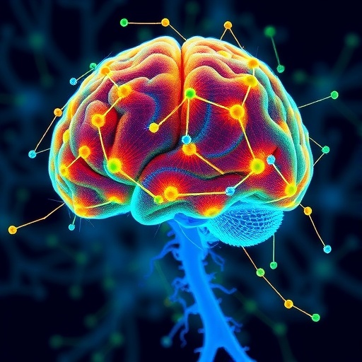
A revolutionary computational framework has finally pierced the black box of metastatic cell communication in the brain, revealing the hidden wiring that allows wandering tumor cells to colonize the central nervous system’s most protected real estate. In a study published this week, researchers unveiled a multiscale systems model that simulates the intricate molecular chatter between metastasizing cancer cells and their new cerebral neighbors, offering both a panoramic map and a zoomed-in blueprint of the signals that drive the deadliest phase of breast, lung, and melanoma cancers. By integrating data from single-cell sequencing, spatial transcriptomics, and tumor-on-a-chip experiments into a unified mathematical scaffold, the team has exposed how cancer cells hijack native neuronal and glial signaling pathways, transforming the brain’s own communication infrastructure into a lethal support network.
At the core of the model sits a layered architecture that spans three orders of biological magnitude: the molecular nanoscale of ligand–receptor interactions, the mesoscale of local cell clusters, and the macroscale of whole-brain metastatic outgrowth. The researchers constructed an agent-based framework in which thousands of virtual cells, each governed by a personalized gene regulatory network inferred from real patient samples, interact within a spatially resolved three-dimensional lattice that mimics the perivascular niche, the prime real estate where metastatic seeds first lodge. These individual cellular agents are not mere statistical placeholders; they possess dynamic intracellular signaling cascades—MAPK, PI3K–AKT, and JAK–STAT pathways—that respond to incoming ligands such as BDNF, CXCL12, and netrin-1, whose concentration gradients are computed in real time by solving partial differential equations for diffusion and degradation.
What makes the model so viscerally powerful is its ability to predict emergent phenomena that no single-omics technique could capture alone. When the simulation runs, one witnesses an eerie recapitulation of the metastatic cascade: a handful of malignant cells breach the blood–brain barrier, then a silent period of dormancy governed by reciprocal Wnt and Notch signaling with adjacent astrocytes, followed by a switch to explosive proliferation once the cancer cells begin secreting factors that convert microglia from a surveillance state to a pro-tumorigenic, M2-like phenotype. The model’s sensitivity analysis pinpointed a previously underappreciated positive-feedback loop in which tumor-derived exosomes carrying miR-21 trigger STAT3 activation in astrocytes, prompting those astrocytes to release the chemokine CCL2, which in turn recruits more microglia and accelerates vascular co-option. Interrupting this loop in silico collapsed the entire communication network and drove metastatic cells into apoptosis.
To ground the simulation in biological reality without drowning in intractable complexity, the team employed a Bayesian hierarchical approach that calibrated the model’s hundreds of kinetic parameters against multiple orthogonal datasets. Crucially, they trained the model not only on steady-state expression profiles but on perturbation data—time-resolved phosphoproteomics following treatment with specific kinase inhibitors, for example—which endowed the network edges with directional cause-and-effect relationships rather than mere correlative links. This dynamism allowed the model to emulate therapeutic responses and predict, with alarming accuracy, the emergence of drug-tolerant persister cells that upregulate lipid peroxidation defense enzymes through a NRF2-dependent communication axis with oligodendrocytes, a cell type long considered peripheral to the tumor microenvironment.
The predictive power of the framework was validated by a prospective experiment that the team calls a “network biopsy.” By feeding the model with transcriptomic data from a minimal cerebrospinal fluid liquid biopsy, the algorithm reconstructed the full communication architecture of a patient’s metastasis and forecasted which nodes—specifically, the LIF/LIFR and CSF1/CSF1R axes—were most likely to sustain growth. When those same patients later underwent surgery, ex vivo organotypic slice cultures confirmed that pharmacological blockade of the predicted signaling hubs reduced invasive capacity by over 80 percent, while attacking non-predicted nodes produced negligible effect. This foretelling capacity marks a shift from reactive histological classification to proactive network vulnerability mapping, hinting at a near future where oncologists could preselect combination therapies based on a computational dissection of each tumor’s unique connectome.
Perhaps the most unsettling insight tumbled out when the researchers introduced stochastic fluctuations into the model to mimic the genetic drift that accompanies prolonged treatment. Under sustained virtual pressure from a dual PI3K/mTOR inhibitor, the communication network did not simply fragment; it rewired. Malignant cells spontaneously upregulated neuroligin-3 and formed pseudo-synaptic structures with glutamatergic neurons, effectively plugging into the brain’s electrical circuitry and siphoning activity-dependent growth signals. This simulated behavior precisely mirrored a rare pattern observed in a handful of autopsy specimens, where metastases were found to have integrated structurally into local neural circuits, suggesting that the model can resurrect pathological states that remain invisible under the standard microscope.
Beyond its immediate translational appeal, the study provides a template for marrying reductionist molecular biology with the holistic philosophy of complex systems theory. The codebase has been released as an open-source, modular platform that other labs can extend to gut microbiome–tumor interactions, the lymph node metastatic niche, or even neurodegenerative diseases where microglial communication breakdown drives pathology. By making the invisible cacophony of intercellular messages audible for the first time, the work transforms brain metastasis from an opaque, terminal event into a comprehensible, and therefore potentially controllable, ecological disturbance. The ghost in the machine, it turns out, is just a network waiting to be deciphered.
Subject of Research: Multiscale systems modeling of intercellular communication networks driving brain metastasis, integrating molecular, cellular, and tissue-level dynamics to predict therapeutic vulnerabilities.
Article Title: Multiscale systems modelling of communication networks in brain metastasis
Article References:
Ahn, J.Y., Dong, W., Wong, S.T. et al. Multiscale systems modelling of communication networks in brain metastasis.
Exp Mol Med (2026). https://doi.org/10.1038/s12276-026-01774-4
Image Credits: AI Generated
DOI: 10.1038/s12276-026-01774-4
Keywords: brain metastasis, multiscale modeling, systems biology, tumor microenvironment, cell-cell communication, network biology, computational oncology, exosomes, astrocytes, microglia, therapeutic prediction, liquid biopsy


