


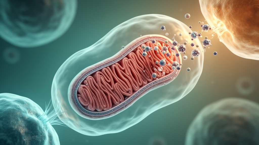
A comprehensive review concludes that nanomaterials could overcome several barriers to mitochondria-targeted therapy by directing cargo to mitochondria, protecting transplanted organelles, increasing intercellular mitochondrial transfer and supporting mitochondrial quality control. The evidence is overwhelmingly preclinical, however, and the review identifies targeting, safety, manufacturing, regulation and long-term integration as unresolved obstacles to clinical translation. Mitochondria generate ATP through oxidative phosphorylation, regulate calcium and reactive oxygen species, participate in cell-death and immune pathways, and maintain their network through fusion, fission, mitophagy and biogenesis.
Disruption of these processes is associated with inherited mitochondrial disorders and acquired diseases affecting energy-intensive organs such as the heart, brain and kidney, as well as metabolic, autoimmune and inflammatory conditions. Current mitochondrial therapeutics broadly follow three strategies: pharmacological modulation of existing mitochondria, transplantation of isolated healthy organelles and exploitation of natural intercellular mitochondrial transfer. Nanomaterials provide a common engineering platform for all three.
Their tunable size, shape and surface chemistry support hierarchical targeting from tissue to cell to organelle, while encapsulation can preserve fragile mitochondria during storage and circulation. Other designs regulate reactive oxygen species, alter energy metabolism, promote mitophagy or biogenesis, remodel inflammatory tissue environments and help mitochondrial cargo escape destructive endolysosomal pathways. Targeting mitochondria remains technically demanding because therapeutic molecules must cross the plasma membrane, escape endosomes and then traverse the outer and inner mitochondrial membranes.
Triphenylphosphonium is widely used because its cationic, lipophilic structure favors accumulation in negatively charged mitochondria, while Szeto–Schiller peptides and mitochondria-penetrating peptides exploit electrostatic and hydrophobic interactions with mitochondrial lipids. Each method has limitations: triphenylphosphonium can act as an uncoupler at concentrations above 10 micromolar, and peptides may be unstable, poorly bioavailable or rapidly degraded in circulation. Nanocarriers can conceal these liabilities, add controlled release and combine tissue- or cell-targeting ligands with organelle-localization signals.
One liposomal formulation achieved 3.7-fold greater mitochondrial accumulation than a triphenylphosphonium-based liposome and eliminated tumors in a 4T1 breast-cancer mouse model, but that finding remains an animal result rather than evidence of human efficacy. The review also describes experimental platforms for mitochondrial gene delivery and editing. Fluorinated lipid nanoparticles and mitochondrial signal sequences are being explored to reach the matrix without relying entirely on membrane potential, which can be impaired in disease.
These demonstrations establish preclinical feasibility, not a validated clinical editing method. Mitochondria can move between cells through tunneling nanotubes, gap junctions, extracellular vesicles, dendritic structures, free release and cell fusion. Such transfer can rescue stressed cells, but it can also spread dysfunctional mitochondria, alter inflammation or support tumors.
In preclinical studies, tumor-derived mitochondria carrying mutant mitochondrial DNA reduced the cytotoxic activity of tumor-infiltrating T cells, whereas mitochondrial transfer from bone marrow–derived mesenchymal stromal cells improved the metabolic fitness and antitumor response of CD8-positive T cells in animal tumor models. Nanomaterials have been used to increase connexin 43 expression, stimulate tunneling nanotubes, construct direct injection devices and engineer donor cells. A photothermal nanoblade delivered isolated mitochondria into recipient cells, while a force-controlled FluidFM system achieved targeted organelle transfer and mitochondrial-DNA propagation during subsequent cell divisions.
Transplanting isolated mitochondria offers a more immediate and controllable energy source, but the organelles rapidly lose activity outside cells, particularly under calcium-rich and oxidative conditions. Extracellular mitochondria can also act as damage-associated molecular patterns, provoking macrophage activation, neutrophil stimulation and, in animal models, increased heart-transplant rejection. Nanomaterials address some of these problems through protective vesicles, artificial membranes, metal–organic frameworks, hydrogels and microneedle patches.
In the reviewed studies, extracellular vesicles preserved mitochondrial structure and function for up to 24 hours under combined calcium and hydrogen-peroxide stress, while microvesicles retained activity for up to 72 hours. Mitochondria enclosed in ZIF-8 remained bioactive for as long as four weeks, and hydrogel or DNA-based systems extended local delivery while also scavenging reactive oxygen species. Several experimental platforms go beyond simple cargo delivery.
Engineered mitochondria have been coupled to neutrophil-hitchhiking systems for sequential targeting in myocardial ischemia–reperfusion injury and to peptides that direct them toward inflamed cardiac tissue. Other composites combine mitochondrial replacement with restoration of mitophagy: attaching healthy mitochondria to nanoparticles carrying Parkin messenger RNA increased respiratory complex I activity from 29.5% in a rotenone-damaged group to 76.5% in a mouse model of complex I deficiency. Biomimetic systems go further, using enzymes, ATP synthase and artificial proton gradients to produce ATP or NADH.
Such artificial mitochondria remain experimental substitutes rather than living organelles, but they offer a cell-free route for addressing energy deficits that has yet to be tested as a clinical treatment. The therapeutic risk can also be double-edged. Mitochondrial transfer is a normal physiological event and may be less immunogenic than transplantation, yet it is usually inefficient, difficult to direct and incompletely understood.
Pathological environments can permit indiscriminate exchange, while mesenchymal stromal cells used as donors raise separate concerns about tumor transformation and off-target differentiation. Some nanomaterials, including cobalt and silver nanoparticles, have demonstrated neurotoxic potential, and nanoplastics have been associated with senescence and cardiac aging through cGAS–STING signaling. Although clinical trials of mitochondrial transplantation are underway in conditions including myocardial infarction, ischemic stroke and inflammatory myopathy, uncertainty remains over dosing, donor–recipient metabolic compatibility, unintended genetic effects and long-term integration.
Translation will therefore require more than a sharper carrier. Future systems must be evaluated for precise tissue and organelle targeting, immune evasion, stimuli-responsive release, stability and safety, as well as reproducible manufacturing. Most of the evidence highlighted in the review comes from conventional cultures and animal models; organoids could provide more physiologically relevant assessments of efficacy and safety.
Artificial-intelligence-assisted formulation and real-time in vivo imaging are proposed as tools for optimizing these designs, but they do not substitute for long-term studies. The review’s central conclusion is promising but conditional: nanomaterials could overcome several barriers that currently restrict mitochondrial therapies, yet reliable clinical benefit will depend on integrated work across materials science, mitochondrial biology, toxicology, regulation and scalable biomanufacturing.
Subject of Research: Nanomaterial-based strategies for mitochondrial targeting, transplantation, intercellular transfer and gene delivery
Article Title: Mitochondria‐Targeted Nanotherapeutics: A Promising Strategy in Modulating Mitochondrial Function, Transfer, and Transplantation
Article References: Su, Y., Gong, Z., Mu, J., Huang, L., Yang, P., Zhang, C., Zhao, L., Yang, L., Deng, J., & Liao, X. (2026). Mitochondria‐Targeted Nanotherapeutics: A Promising Strategy in Modulating Mitochondrial Function, Transfer, and Transplantation. Advanced Science, 13(58), Article e77713. https://doi.org/10.1002/advs.77713
Image Credits: AI Generated
DOI: 10.1002/advs.77713
Keywords: mitochondrial therapy, nanomedicine, mitochondrial transplantation, intercellular transfer, nanocarriers, mitochondrial targeting, mitochondrial DNA, mitophagy, extracellular vesicles, biomimetic nanomaterials, Mitochondria, Targeted
Cite Scienmag News
Drew Townsend. (October 20, 2026). Mitochondria‐Targeted Nanotherapeutics: A Promising Strategy in Modulating Mitochondrial Function, Transfer, and Transplantation. Scienmag. https://scienmag.com/mitochondria%e2%80%90targeted-nanotherapeutics-a-promising-strategy-in-modulating-mitochondrial-function-transfer-and-transplantation/
Drew Townsend. "Mitochondria‐Targeted Nanotherapeutics: A Promising Strategy in Modulating Mitochondrial Function, Transfer, and Transplantation." Scienmag, 20 October 2026, https://scienmag.com/mitochondria%e2%80%90targeted-nanotherapeutics-a-promising-strategy-in-modulating-mitochondrial-function-transfer-and-transplantation/. Accessed 20 October 2026.
Drew Townsend. "Mitochondria‐Targeted Nanotherapeutics: A Promising Strategy in Modulating Mitochondrial Function, Transfer, and Transplantation." Scienmag. October 20, 2026. https://scienmag.com/mitochondria%e2%80%90targeted-nanotherapeutics-a-promising-strategy-in-modulating-mitochondrial-function-transfer-and-transplantation/


