


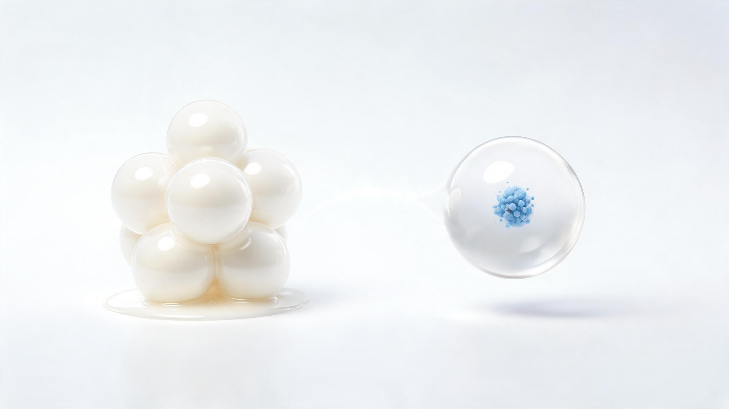
Milk may be more than a source of calories, proteins and minerals: it also carries microscopic parcels that researchers are studying as potential delivery vehicles for medicines and nutraceuticals. A recent review examines milk-derived extracellular vesicles, or mEVs, naturally occurring particles enclosed by lipid membranes and typically measuring tens to hundreds of nanometres across. Secreted mainly by mammary epithelial and immune cells, these vesicles transport proteins, lipids, metabolites, messenger RNAs and microRNAs between cells. Their biological role in milk is linked to communication, immune development and metabolic regulation, while their physical structure has attracted interest in nanomedicine. Unlike many synthetic nanoparticles, mEVs arise from a food-associated biological system and may be produced from abundant milk supplies. The review presents them as a possible bridge between nutrition and therapeutics, while emphasizing that most evidence remains preclinical and that major manufacturing, safety and regulatory questions must be resolved before broad clinical use.
The appeal of mEVs begins with their membrane. Cholesterol, sphingomyelin and ceramides help create a relatively robust lipid bilayer that shields internal cargo from environmental damage. Studies summarized in the review indicate that milk vesicles can remain intact under simulated gastrointestinal conditions and protect RNA from digestive enzymes such as ribonucleases. After oral administration, vesicles may interact with intestinal epithelial cells through endocytosis, membrane fusion or receptor-mediated uptake, allowing their contents to enter recipient cells. Some experimental work also suggests that milk vesicles or their cargo can reach tissues beyond the gut, including the brain. This possibility is particularly important because the blood-brain barrier restricts many therapeutic molecules. However, crossing that barrier in animal or cellular models does not establish effective delivery in people. Biodistribution depends on vesicle size, surface proteins, cargo, dose, species of origin, processing conditions and administration route. The review therefore treats gastrointestinal stability and barrier transport as promising properties, not guarantees of therapeutic performance.
Milk vesicles are not a single uniform material. The broader extracellular-vesicle population includes exosomes, microvesicles and apoptotic bodies, which differ in size and how they form. Exosomes develop inside multivesicular bodies when endosomal membranes bud inward to create intraluminal vesicles; multivesicular bodies can then fuse with the plasma membrane and release them. Microvesicles form by outward budding of the cell surface, involving calcium-dependent cytoskeletal changes and redistribution of membrane lipids. In milk, the vesicle population is shaped by the animal species, lactation stage, maternal physiology, diet and health status. Bovine, human, goat, camel, porcine and equine milk can therefore contain different mixtures of proteins and regulatory RNAs. Commonly measured surface or intracellular markers include CD9, CD63, CD81, TSG101 and Alix, but marker detection alone does not define purity or biological function. The review calls for multi-method characterization that combines particle sizing, microscopy, protein analysis, RNA profiling and functional testing.
Obtaining clean vesicles from milk is technically difficult because milk is a complex mixture containing casein micelles, soluble proteins, fat globules and other particles with overlapping physical properties. Differential ultracentrifugation remains widely used, separating material according to size and density, but it can be slow and may promote aggregation or structural damage. Ultrafiltration and polyethylene-glycol precipitation are easier to scale, yet they can recover non-vesicular contaminants. Size-exclusion chromatography separates particles by hydrodynamic size and is often combined with other methods to improve purity. Immunoaffinity capture can select vesicles carrying particular surface markers, although it may reduce recovery and exclude biologically relevant subpopulations. Emerging microfluidic and immunomagnetic systems could enable automated processing of small volumes, while tangential-flow filtration combined with chromatography offers a possible route toward larger-scale production. Across all approaches, researchers need consistent measurements of particle number, size distribution, morphology, membrane integrity, cargo and contaminating milk proteins.
As delivery systems, mEVs could carry molecules that otherwise degrade quickly or dissolve poorly. The review describes experimental loading with polyphenols such as curcumin, resveratrol, quercetin and epigallocatechin gallate, as well as chemotherapeutic compounds including paclitaxel and doxorubicin. Encapsulation may improve aqueous dispersal, protect cargo during digestion and increase contact with intestinal tissues. Vesicles have also been investigated for transporting small interfering RNA, microRNA, messenger RNA, peptides and proteins. In principle, this creates a dual-purpose platform: the vesicle’s native cargo may influence recipient cells, while an added therapeutic molecule supplies a designed activity. Surface engineering could further attach targeting ligands or alter tissue distribution. Yet loading is not straightforward. Passive incubation, membrane permeabilization and other approaches can produce different encapsulation efficiencies and may damage the vesicle. A useful product would require reproducible cargo content, predictable release kinetics and evidence that the loaded molecule reaches the intended tissue at a clinically meaningful dose.
The biological effects reported across models are broad but uneven. In intestinal systems, milk vesicles have been associated with stronger tight junctions, including proteins such as ZO-1, occludin and claudin-1, and with reduced inflammatory signaling. MicroRNAs including miR-148a, miR-21, miR-30a and miR-146b may influence pathways involving NF-κB, Toll-like receptors, DNA methylation and the NLRP3 inflammasome. In cell and animal studies, these mechanisms have been linked to lower inflammatory cytokines, improved barrier function and protection from oxidative stress. Other experiments report effects on macrophage polarization, with some mEV preparations encouraging an anti-inflammatory state. Researchers have also examined bone, liver, heart, lung and pancreatic applications. Milk vesicles have been tested in models of colitis, metabolic dysfunction, fibrosis, osteoporosis, vascular injury and pulmonary inflammation. These findings suggest multiple possible mechanisms, including direct cargo transfer, modulation of gut microbiota and communication along the gut-liver or gut-heart axes, but they do not demonstrate that drinking milk or consuming an unstandardized vesicle preparation treats disease.
Several findings illustrate why careful interpretation is essential. In mice, orally administered milk vesicles have been reported to cross the blood-brain barrier, increase hippocampal dendritic complexity and improve selected cognitive or motor outcomes. Other studies have found changes in microglial DNA-methylation machinery or neuronal survival in cellular models. At the same time, neurological results vary with dose, species, metabolic context and experimental design. A separate line of research has raised hypotheses about interactions between milk exosomes and excessive galactose exposure, while other work found that aging had stronger effects on rat brain lipid profiles and cognition than an extracellular-vesicle-rich supplement. Lung studies likewise include contrasting observations: some mEV preparations protect epithelial barriers or deliver anti-fibrotic compounds, whereas bovine vesicles increased inflammatory macrophage polarization in mice exposed to agricultural dust. In cancer research, vesicles have delivered drugs and gene regulators to tumour models, but one study reported that oral bovine milk vesicles slowed primary-tumour growth while accelerating metastasis. Such results make clear that mEVs are biologically active, context-dependent materials rather than universally beneficial particles.
Translation will depend on proving safety and manufacturing consistency as much as on demonstrating biological activity. Milk origin does not automatically eliminate risk. Preparations can retain caseins, beta-lactoglobulin and other proteins that may trigger reactions in people with milk allergy. Repeated exposure also requires assessment of immune activation, liver and kidney function, oxidative stress, tissue distribution and delayed toxicity. Thermal processing can reduce vesicle yield or disrupt structure, complicating the relationship between fresh milk, pasteurized products and purified formulations. Regulators will also need to determine whether a given product is a food, supplement, biologic, nanomedicine or combination product, since each category carries different requirements. The review points toward standardized isolation protocols, validated vesicle markers, sensitive contaminant testing, single-vesicle and multi-omic analysis, stable storage methods and Good Manufacturing Practice production. Human pharmacokinetic and clinical studies will be decisive. For now, milk-derived extracellular vesicles represent a promising natural nanocarrier platform whose future rests on converting intriguing laboratory observations into reproducible, well-controlled and demonstrably safe interventions.
A further advantage of mEVs is that their value may extend beyond their role as passive containers. Their membranes carry naturally occurring adhesion molecules, tetraspanins and transport-related components that can influence how vesicles are recognized, internalized and distributed. This biological interface distinguishes them from liposomes and polymeric nanoparticles, whose composition can be tuned with considerable precision but generally requires deliberate surface engineering to achieve comparable interactions with cells. The contrast is not absolute: synthetic systems offer stronger control over particle uniformity, drug loading and release kinetics, while mEVs offer a more physiologically integrated membrane and a potentially favorable safety profile. Hybrid designs that combine EV membranes with synthetic cores therefore represent one strategy for balancing biological compatibility with manufacturing control.
The native cargo also complicates how mEV products should be designed and evaluated. A preparation intended to deliver an added drug or RNA may simultaneously contain endogenous proteins, lipids, microRNAs and metabolites capable of altering immune or metabolic responses. Those constituents could contribute to efficacy, but they could also vary with animal species, lactation conditions, feed, health status and processing history. Consequently, measuring total particle concentration is insufficient for comparing products. Functional potency assays will need to establish whether a defined preparation produces a reproducible cellular response, while molecular profiling can help identify which cargo components are retained, enriched or lost during isolation and loading. This is especially important when the desired activity depends on cooperation between the vesicle membrane and its internal contents rather than on a single therapeutic molecule.
Manufacturing scale is promising but should not be confused with readiness for routine clinical use. Milk provides a comparatively accessible starting material, and the review describes ultracentrifugation, size-exclusion chromatography and precipitation methods as established approaches, with tangential-flow and related technologies offering routes toward process intensification. At larger scale, however, purification must preserve membrane integrity while removing abundant non-vesicular milk constituents and maintaining consistent particle and cargo characteristics. The field has reached early translational milestones, including a reported first clinical trial involving mEV-based formulations for RNA therapeutics and anticancer agents, but such studies are only an initial test of feasibility. Results from carefully controlled human investigations will need to define dose, absorption, biodistribution, immune effects and clinically meaningful benefit before the farm-to-pharmacy concept can support approved interventions.
Subject of Research: Milk-derived extracellular vesicles as nutritional and therapeutic nanocarriers
Article Title: Milk-derived extracellular vesicles: nutritional significance, nano-delivery potential, and emerging therapeutic applications – an updated review
Article References: Wang, S., Shaukat, A., Al-Rasheed, M., Tareen, A. M., Arain, M. A., & Luo, C. (2026). Milk-derived extracellular vesicles: nutritional significance, nano-delivery potential, and emerging therapeutic applications – an updated review. Food Science of Animal Resources, 46(1), Article 96. https://doi.org/10.1007/s44463-026-00104-6
Image Credits: AI Generated
DOI: 10.1007/s44463-026-00104-6
Keywords: extracellular vesicles, milk nanocarriers, nutraceutical delivery, drug delivery, microRNAs, precision medicine, gut health, nanomedicine, Milk-derived, extracellular, vesicles, nutritional
Cite Scienmag News
Louis Brooks. (August 29, 2026). Milk Vesicles Could Bridge Nutrition and Precision Drug Delivery. Scienmag. https://scienmag.com/milk-vesicles-could-bridge-nutrition-and-precision-drug-delivery/
Louis Brooks. "Milk Vesicles Could Bridge Nutrition and Precision Drug Delivery." Scienmag, 29 August 2026, https://scienmag.com/milk-vesicles-could-bridge-nutrition-and-precision-drug-delivery/. Accessed 5 October 2026.
Louis Brooks. "Milk Vesicles Could Bridge Nutrition and Precision Drug Delivery." Scienmag. August 29, 2026. https://scienmag.com/milk-vesicles-could-bridge-nutrition-and-precision-drug-delivery/


