


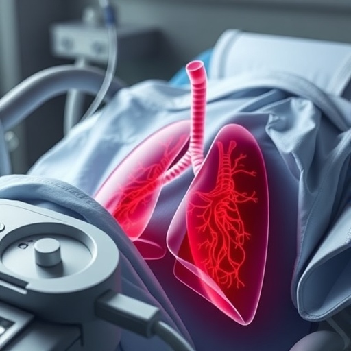
Acute respiratory distress syndrome, or ARDS, is one of the most dangerous complications that can follow severe trauma, and a new review in Intensive Care Medicine argues that injured patients require a more tailored approach than the label “standard ARDS” might suggest. The condition develops when widespread inflammation damages the thin barrier between lung air spaces and blood vessels. Fluid then leaks into the alveoli, the microscopic sacs where oxygen enters the bloodstream, while portions of the lung collapse or become filled with inflammatory material. The result is severe hypoxemia, increased work of breathing and, in many cases, the need for mechanical ventilation. Trauma-related ARDS is especially common after major blunt chest injury, pulmonary contusion and polytrauma, yet the evidence guiding its treatment has largely been borrowed from studies of medical illnesses such as pneumonia and sepsis.
The review describes trauma-related ARDS as a biological “multi-hit” process. The first hit is the physical injury itself: fractured ribs, bruised lung tissue, pneumothorax, hemothorax or damage to other organs. A second hit emerges as the body mounts an intense inflammatory response. Injured cells release damage-associated molecular patterns, while immune cells produce cytokines including tumor necrosis factor alpha, interleukin-1 beta, interleukin-6 and interleukin-8. These signals activate the vascular endothelium and attract neutrophils into the lungs. Neutrophils can intensify tissue damage by releasing proteases, reactive oxygen species and extracellular traps designed to capture pathogens. The alveolar-capillary barrier becomes porous, allowing protein-rich fluid to flood the air spaces. Additional insults—massive transfusion, infection, sepsis, fluid overload, fat embolism or major surgery—may act as third hits, pushing a partially injured lung into full respiratory failure.
Although trauma represents only a small fraction of ARDS cases across intensive care units, the risk rises sharply in patients with serious thoracic injuries. In one French cohort of severely injured adults with significant pulmonary contusion, more than three-quarters required intubation and over one-third developed ARDS. Other studies have reported ARDS in approximately 60 percent of intensive-care patients with major thoracic trauma. Mortality is often lower than in medical ARDS, largely because trauma patients tend to be younger and may have fewer chronic diseases, but the condition remains highly consequential. Contemporary data also suggest that mortality among trauma patients with ARDS has increased over time, despite a declining incidence. Hemorrhagic shock, traumatic brain injury, anticoagulant use, kidney injury and severe sepsis were among the factors associated with death. Risk tools such as the Thoracic Trauma Severity score can combine age, rib fractures, contusion extent, pleural injury and oxygenation to identify patients likely to deteriorate.
Imaging is central because lung contusions frequently worsen during the first 24 to 72 hours, sometimes before conventional chest radiography reveals the full extent of damage. Portable chest X-rays remain valuable for rapidly identifying immediately dangerous problems, checking tubes and drains, and monitoring unstable patients, but they can miss early edema and small or deep injuries. Lung ultrasound can be performed repeatedly at the bedside and can detect pneumothorax, hemothorax and peripheral pulmonary contusions more rapidly than an X-ray in many situations. Ultrasound patterns of alveolar-interstitial injury may also help predict which patients will develop ARDS within the next three days. When transport is safe, computed tomography provides a more detailed map of parenchymal injury, vascular damage, pleural collections and rib fractures. Contusions involving more than roughly one-fifth to one-quarter of total lung volume have been associated with increased risks of ARDS and pneumonia, although imaging findings must always be interpreted alongside clinical physiology.
The review emphasizes that respiratory support should be protective but carefully individualized. In patients who are awake, cooperative and not severely hypoxemic, high-flow nasal oxygen or non-invasive ventilation may reduce the need for intubation, particularly when effective analgesia allows deep breathing and coughing. However, non-invasive support must not become a reason to delay intubation in a deteriorating patient. Excessive inspiratory effort can generate large swings in transpulmonary pressure, a phenomenon known as patient self-inflicted lung injury. The ROX index, which relates oxygen saturation and inspired oxygen concentration to respiratory rate, may help identify success or failure, but trauma patients appear to require higher thresholds than patients with ordinary medical respiratory failure. In selected cases, helmet-based non-invasive ventilation may improve tolerance, and limited sedation with dexmedetomidine has been investigated to help patients complete longer treatment sessions without substantially suppressing respiratory drive.
Once invasive ventilation is necessary, the principles used in general ARDS remain the foundation: low tidal volumes based on predicted body weight, limitation of plateau pressure and close attention to driving pressure, the difference between plateau pressure and positive end-expiratory pressure. Trauma creates an additional challenge because the lung is often highly uneven. Dense focal contusions can exist beside relatively healthy or overinflated regions, meaning that a single ventilator setting may recruit injured areas while overstretching healthier tissue. PEEP should therefore reflect lung morphology and recruitability rather than being increased automatically. Higher PEEP may benefit diffuse, recruitable disease but can worsen overdistension in focal injury. Prone positioning can improve oxygenation and survival in trauma-associated ARDS, although unstable spinal injuries, fractures and surgical wounds may make it hazardous. Alternating lateral positioning has also been explored. For refractory hypoxemia, venovenous extracorporeal membrane oxygenation may be considered in specialized centers, with anticoagulation adjusted to the patient’s bleeding risk.
Fluid and circulatory management create another delicate balance. A trauma patient in hemorrhagic shock needs rapid restoration of blood volume and oxygen-carrying capacity, but excessive crystalloid or blood-product administration can intensify pulmonary edema and inflammation. Before bleeding is controlled, permissive hypotension with a systolic blood pressure around 80 to 90 millimeters of mercury may be appropriate in patients without brain injury, limiting the pressure that drives further hemorrhage. The strategy changes when traumatic brain injury is present, because inadequate cerebral perfusion can worsen neurological damage. After initial resuscitation and hemodynamic stabilization, the evidence favors avoiding unnecessary fluid accumulation. A conservative approach can improve oxygenation and shorten ventilation, while dynamic assessment of fluid responsiveness, lung water, cardiac function and perfusion helps determine whether another bolus is likely to help or harm. During recovery, carefully monitored fluid removal may reduce pulmonary congestion and support liberation from the ventilator.
Pain control is presented not as a comfort measure but as a form of respiratory therapy. Rib fractures make every breath painful, encouraging shallow ventilation and suppressing cough. Secretions then accumulate, raising the risk of atelectasis, mucus plugging, pneumonia and respiratory failure. The PIC score, which combines pain, inspiratory effort and cough effectiveness, attempts to measure how injury is affecting breathing rather than simply asking how severe the pain feels. Multimodal treatment may include paracetamol, anti-inflammatory drugs, carefully selected opioids and ketamine, which can provide analgesia while preserving respiratory drive. Regional techniques, including nerve blocks and other targeted approaches, can improve breathing and physiotherapy while reducing systemic opioid exposure, but must be chosen according to coagulation status, anatomy and hemodynamic stability. In patients with multiple rib fractures, early surgical stabilization may also improve chest-wall mechanics, reduce pain and facilitate weaning, although randomized evidence remains mixed and the procedure should not be applied automatically.
Surgical timing can influence lung outcomes beyond the chest itself. Early stabilization of femoral and pelvic fractures may reduce the continuing release of inflammatory mediators and fat particles into the circulation, potentially lowering the risk of ARDS and pneumonia. Damage-control surgery may be preferable when shock, hypothermia, acidosis or coagulopathy make prolonged definitive procedures dangerous. For the chest wall, rib fixation is most often considered when respiratory compromise, flail chest, prolonged ventilation or uncontrolled pain persists despite optimized treatment. Some observational studies associate early fixation with fewer pulmonary complications, but other controlled research has found no improvement in ventilator-free days among selected patients without clinical flail chest. The review therefore portrays surgery as part of a multidisciplinary decision rather than a universal solution. Its final warning concerns patients with simultaneous traumatic brain and lung injury: oxygen and carbon dioxide targets, PEEP, prone positioning and intracranial-pressure treatments must be adjusted together, because improving one organ can sometimes compromise the other. The authors conclude that trauma-specific trials are urgently needed to define biological subtypes, predict non-invasive support failure and determine which combinations of ventilation, analgesia, fluid control and early surgery truly improve survival.
Cite Scienmag News
Ophelia Keating. (August 26, 2026). Managing Acute Respiratory Distress Syndrome in Trauma Patients. Scienmag. https://scienmag.com/managing-acute-respiratory-distress-syndrome-in-trauma-patients/
Ophelia Keating. "Managing Acute Respiratory Distress Syndrome in Trauma Patients." Scienmag, 26 August 2026, https://scienmag.com/managing-acute-respiratory-distress-syndrome-in-trauma-patients/. Accessed 3 September 2026.
Ophelia Keating. "Managing Acute Respiratory Distress Syndrome in Trauma Patients." Scienmag. August 26, 2026. https://scienmag.com/managing-acute-respiratory-distress-syndrome-in-trauma-patients/


