


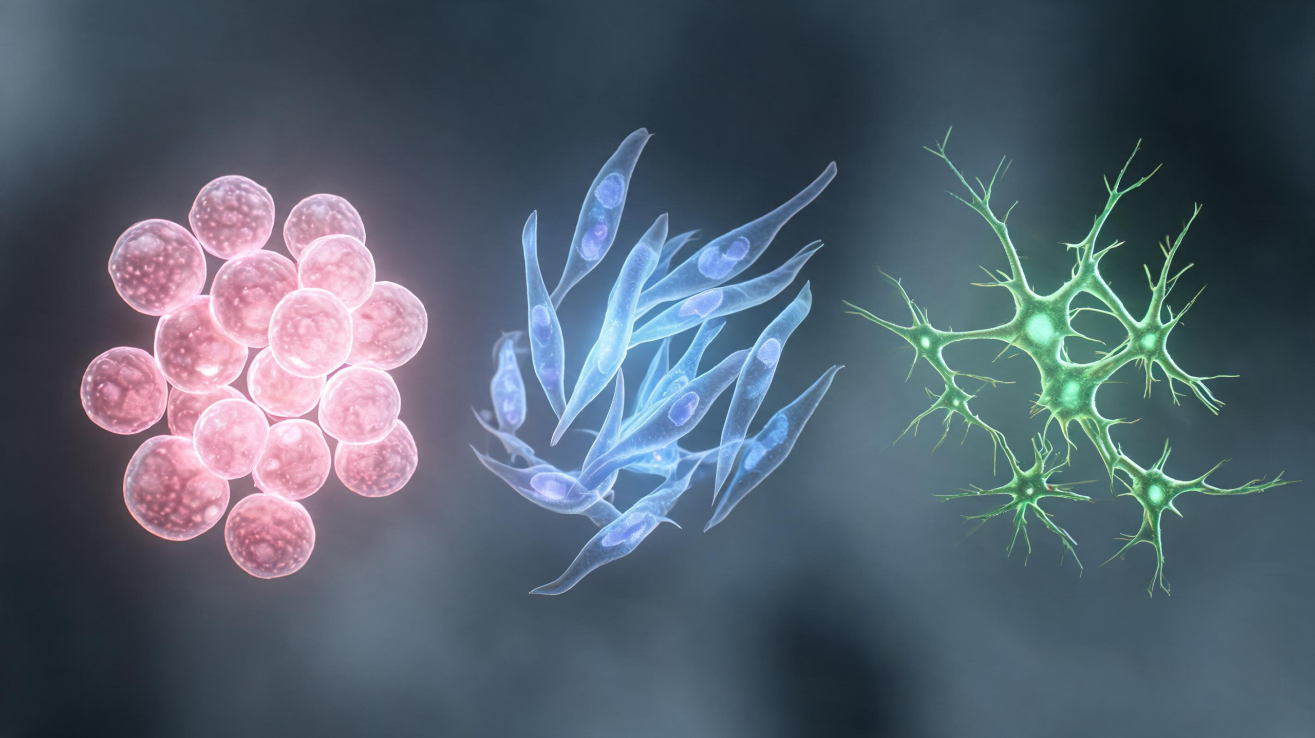
Cells do not become specialized in a single instant. During development, they gradually change their molecular programs, position, shape, polarity and behavior before reaching a mature identity. A study in Molecular Systems Biology highlights a way to follow that process as it unfolds, using live imaging and computational analysis of cell morphology. The approach, known as morphodynamics, treats a cell’s changing physical features as information about its developmental state. Julia Dorr and Brian J. Mitchell describe how recent work by Tolonen and colleagues used this strategy to predict the eventual fates of individual cells in a developing Xenopus mucociliary epithelium. The work addresses a central limitation of modern cell biology: many widely used measurements record molecular states at selected time points, while differentiation is a continuous and dynamic process. By tracking cells through time, researchers can examine not only what genes are active, but also how cells move, rearrange their internal geometry and interact with neighboring cells as their destinies emerge. The result is a computational framework that begins to connect cell appearance and behavior with the formation of functioning tissue.
Much of the current understanding of cell fate comes from single-cell molecular measurements, particularly RNA sequencing. These methods can provide detailed profiles of gene expression and reveal regulatory networks associated with distinct cell states. Yet each measurement is usually a snapshot: it captures the molecular condition of a cell after the sample has been collected, rather than continuously observing the transition that produced it. Computational methods such as pseudotime analysis can arrange cells along an inferred developmental trajectory, but an inferred sequence is not the same as a direct record of change. Morphodynamics offers a complementary perspective by measuring features that can be observed repeatedly in living cells. Those features may include cell area, shape, movement, position within a tissue, nuclear geometry and the relationship between cellular structures. In principle, a time-resolved record of these variables can reveal transitional states that molecular sampling misses. The objective is not to replace molecular profiling, but to add the physical and behavioral dimension needed to understand how cell states are established in real tissues.
Tolonen and colleagues selected the Xenopus mucociliary epithelium because it differentiates rapidly and produces a complex, multilayered tissue. The model begins with an ectodermal cap that can be excised from an embryo and attached to a fibronectin-coated glass-bottom dish. Under culture conditions, the tissue proceeds through much of its differentiation program while remaining sufficiently thin for high-resolution, long-term live imaging. Over approximately 22 hours, it develops into a mature bilayer containing several specialized cell types. These include multiciliated cells, which help move material across an epithelial surface; small secretory cells; ionocytes; goblet cells; and basal stem cells. The cell types differ in their morphology and movement trajectories, creating observable physical signatures that can be measured during development. Because the explant reproduces important features of mucociliary epithelial development and resembles aspects of mammalian airway epithelium, it provides a tractable system for studying how cell behavior contributes to tissue organization.
To follow individual cells, the researchers used embryos injected with fluorescent markers labeling nuclei and cell membranes. Live imaging then captured the developing epithelium in three dimensions, while segmentation and tracking tools converted the image sequence into individual cell trajectories. Segmentation assigns image pixels or voxels to a particular cell, creating a digital mask that defines its boundaries. Tracking links those masks across successive frames, allowing researchers to estimate how each cell moves and changes over time. The analysis faced practical challenges. Cell shapes varied, the tissue was compact, and the available resolution along the imaging axis was limited. Membrane boundaries could therefore be difficult to identify consistently. Nuclear labeling provided a more reliable anchor, enabling accurate lineage tracking even when the surrounding cell geometry was ambiguous. From these trajectories, the team extracted morphometric and dynamic measurements and used them to define a morphodynamic state for each cell. This concept parallels a molecular state defined by gene expression, but it is based on physical features and behavior recorded in living tissue.
The first analysis produced an instructive negative result. When individual cellular features were considered without broader lineage information, the cells did not form sharply separated clusters corresponding to their eventual identities. The absence of clear clusters suggests that the relevant differences in this tissue are subtle rather than dramatic. Epithelial cells are also subject to physical constraints: they must pack together, share boundaries and maintain tissue integrity, which can make distinct cell types look similar at particular moments. Differentiation may therefore be encoded not in one conspicuous feature, but in combinations of modest changes distributed across time. To address this problem, the researchers turned to supervised machine-learning models. They generated a ground-truth dataset by fixing and immunostaining tissues at the endpoint, assigning final cell identities and then tracing those cells backward through their recorded lineages. This provided the models with known outcomes against which earlier morphodynamic patterns could be tested, transforming subtle physical trends into measurable associations with fate.
The supervised analysis used multivariate, multiclass prediction methods, including XGBoost and multinomial logistic regression implemented with scikit-learn. Rather than asking whether one measurement alone identified a cell type, these models evaluated combinations of features and their contribution to classification. XGBoost, an ensemble method based on decision-tree boosting, produced a mean cell-fate prediction accuracy of approximately 80 percent in the reported analysis. The most influential feature was the cell’s position along the Z axis. That result is biologically plausible because certain differentiated cell types occupy the apical surface of the multilayered epithelium. The model also identified the offset between nuclear and membrane centroids as informative. This measurement can reflect changes in cell polarity, shape and spatial organization during morphogenetic events such as radial intercalation, when cells move between tissue layers or rearrange relative to their neighbors. These signals were not necessarily strong enough to identify fate in isolation. Their predictive value emerged when the model considered several measurements together and interpreted them in the context of a cell’s lineage.
The findings illustrate why time-resolved phenomics could become an important partner to single-cell omics. Molecular data can show which genes and regulatory pathways are associated with a transition, whereas morphodynamic data can reveal when a cell changes position, how it reshapes itself and whether its movements are coordinated with those of nearby cells. Such information is especially relevant in epithelia, where fate is linked to tissue architecture, mechanical forces and collective behavior. A cell’s final identity may depend partly on the physical environment it experiences as it moves through a crowded, curved or multilayered tissue. Live imaging preserves this context, while computational pipelines make it possible to quantify many cells across extended developmental windows. Previous work has shown that single-cell phenomics can expose behavioral and mechanical heterogeneity during tissue remodeling. The current analysis extends that idea by showing that dynamic physical measurements can be scaled into a predictive framework resembling an omics workflow, even when individual features are relatively weak and cell states change continuously.
Several limitations remain before morphodynamics can provide a complete account of cell fate. The imaging system depends on fluorescent labeling, reliable segmentation and sufficient spatial and temporal resolution, and the authors note that variable morphology and limited Z resolution can affect the resulting masks. Prediction accuracy also depends on the quality and scope of the ground-truth dataset used for training. A model trained in one developmental system may not transfer directly to another tissue, species or disease state. Future studies could combine live morphodynamic measurements with molecular profiling of the same cells or closely matched lineages. Such integration may identify the precise time points at which physical changes coincide with decisive regulatory events. The approach could also help establish baseline patterns for biomedical phenotyping and early disease detection. In cancer research, for example, a detailed understanding of how normal cells change shape, position and behavior during differentiation could make it easier to recognize abnormal departures from that program. The broader significance is that cell fate may be read not only in molecular snapshots, but also in the evolving geometry and motion of living cells.
An important conceptual shift in this work is the treatment of morphology as a state variable rather than merely an endpoint description. A cell’s location, geometry and movement can be recorded repeatedly, preserving the order in which changes occur. This makes it possible to ask whether a physical feature precedes the appearance of a mature marker, rather than simply correlating the two after differentiation has finished. The distinction is especially valuable for identifying transition windows in which a cell may still be responsive to its environment or susceptible to developmental perturbation.
The study also shows why lineage information is central to interpreting phenotypic measurements. Cells sharing a tissue compartment may appear similar at one time point even when their later outcomes diverge. Conversely, the same feature may have different implications depending on where a cell came from and how it has moved. Linking measurements across a trajectory therefore supplies context that a collection of unrelated images cannot provide. Endpoint immunostaining served as the reference for assigning outcomes, while the preceding live record supplied the evidence used for prediction. This combination connects retrospective identity measurements with prospective dynamics without assuming that every visible difference is fate-determining.
Prediction should nevertheless be distinguished from mechanism. An informative feature, such as apical position or nuclear–membrane displacement, may report a process that accompanies fate commitment without causing it. The predictive pipeline can reveal when and where such associations occur, but perturbation experiments would be needed to test their functional importance. The framework could consequently serve as a way to prioritize developmental time points, cellular behaviors or physical transitions for experimental intervention. In this sense, morphodynamic analysis is not only a classification strategy: it can organize the complex sequence of events that connects progenitor behavior to the architecture of a differentiated epithelium.
Subject of Research: Using live-cell morphodynamics to predict cell fate during epithelial differentiation
Article Title: Tracking cell fate through morphodynamics
Article References: Dorr, J., & Mitchell, B. J. (2026). Tracking cell fate through morphodynamics. Molecular Systems Biology. https://doi.org/10.1038/s44320-026-00244-3
Image Credits: AI Generated
DOI: 10.1038/s44320-026-00244-3
Keywords: cell fate, morphodynamics, live-cell imaging, epithelial development, Xenopus, single-cell phenomics, machine learning, cell differentiation, Tracking, cell, fate, scientific research
Cite Scienmag News
Drew Townsend. (August 28, 2026). Live Cell Shapes Reveal How Tissues Choose Their Final Identities. Scienmag. https://scienmag.com/live-cell-shapes-reveal-how-tissues-choose-their-final-identities/
Drew Townsend. "Live Cell Shapes Reveal How Tissues Choose Their Final Identities." Scienmag, 28 August 2026, https://scienmag.com/live-cell-shapes-reveal-how-tissues-choose-their-final-identities/. Accessed 5 October 2026.
Drew Townsend. "Live Cell Shapes Reveal How Tissues Choose Their Final Identities." Scienmag. August 28, 2026. https://scienmag.com/live-cell-shapes-reveal-how-tissues-choose-their-final-identities/


