A molecule extracted from the leaves of a wild Himalayan plant can spot hypochlorite, the reactive anion behind household bleach and water disinfection, with a simple color change visible to the naked eye. The compound, xanthotoxin, was isolated from Prangos pabularia, an aromatic member of the carrot family that grows on the stony slopes of Drass in the Kargil district of Ladakh, India. In a study published in Results in Chemistry, a research team based in India reports that this naturally occurring furanocoumarin acts as a selective, reversible and rapid colorimetric sensor for hypochlorite in acetonitrile, reaching a detection limit of 57 micromolar and producing a full visual response within roughly one minute.
The appeal of the approach lies in its simplicity. Most existing colorimetric probes for hypochlorite are synthetic organic molecules that demand multistep laboratory syntheses and generate chemical waste along the way. By contrast, xanthotoxin is a ready-made natural product, obtained here by shade-drying the collected leaves for a month, grinding them into a powder, macerating the powder in methanol for fifteen days, and then purifying the crude extract by silica gel column chromatography with a hexane-to-ethyl acetate gradient. The procedure yielded 40 milligrams of xanthotoxin as a white solid, whose identity was confirmed by nuclear magnetic resonance spectroscopy, infrared spectroscopy and high-resolution mass spectrometry.
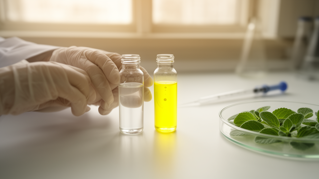
When the researchers dissolved the compound in acetonitrile and added aqueous solutions of ten different anions, only hypochlorite triggered a reaction. Fluoride, chloride, bromide, iodide, sulfate, nitrate, nitrite, hydrogen sulfate and hydrogen phosphate all left the solution untouched, while hypochlorite turned it from colorless to yellow immediately. Ultraviolet-visible spectroscopy revealed what was happening at the molecular level: the sensor’s original absorption peak at 296 nanometers gave way to a new band at 358 nanometers, a bathochromic shift of 89 nanometers, and an isosbestic point near 325 nanometers indicated a clean equilibrium between the free sensor and its hypochlorite-bound form.
The response was also strikingly reversible. Adding a protic acid such as acetic acid or hydrochloric acid to the yellow solution wiped out the color at once, restoring the original colorless state. This acid-driven reset suggests the underlying chemistry is an equilibrium rather than a destructive reaction, which opens the door to reusing the same sensing solution. A Job’s plot analysis, in which the mole fractions of sensor and hypochlorite were varied while their total concentration was held constant, showed that the two species associate in a one-to-one ratio.
Timing experiments sharpened the picture of the mechanism. When the absorbance of a sensor solution containing 0.1 millimolar hypochlorite was monitored over seconds, the signal climbed for about 60 seconds and then plateaued, a pattern the authors interpret as evidence for a reaction-based process rather than simple physical binding. Based on this and the spectroscopic evidence, they propose that hypochlorite attacks the electron-deficient carbonyl carbon of the coumarin ring, cleaving the lactone ester bond and reversibly opening the ring to form a hypochlorite adduct. High-resolution mass spectrometry of the reacted mixture detected a species whose observed mass matched the calculated exact mass of 267.0066 for the proposed adduct, supporting the ring-opened intermediate.
The ring-opening chemistry also explains the color. Closing the lactone keeps the molecule’s pi-electron system fully conjugated; opening it disrupts that conjugation and shifts the absorption into the visible range, producing the yellow hue. Under acidic conditions, hypochlorite is converted predominantly to hypochlorous acid, which favors intramolecular cyclization and restores the closed coumarin ring, returning the electronic conjugation and the colorless appearance. Consistent with this mechanism, pH tests showed a strong absorption band near 358 nanometers at pH values of 7.9 and above, while acidic conditions below pH 6.5 produced no significant spectral change, presumably because the available hypochlorite had been protonated away. The authors conclude that the sensor works best under near-neutral conditions, with HEPES buffer at pH 7.4 providing a suitable medium.
Quantitatively, the sensor performed respectably for a natural-product platform. A calibration curve built from absorbance measurements at 358 nanometer showed good linearity across the tested concentration range, with a correlation coefficient of 0.98 based on three independent measurements. Applying the standard formula LOD = 3σ/S, where σ is the standard deviation of blank readings and S the calibration slope, gave a detection limit of 57 micromolar. The sensor pairs its detection limit with a visible color change requiring no instrument, a sustainable and low-toxicity source molecule, and straightforward reversibility.
The team also characterized the compound’s electronic structure computationally, using density functional theory at the B3LYP/6-31G(d,p) level and time-dependent DFT to simulate the absorption spectrum. In the gas phase, the calculated HOMO-LUMO energy gap was 4.052 electronvolts; adding an acetonitrile solvent model lowered it to 3.956 electronvolts. The experimental optical band gap, estimated from the absorption edge of the ultraviolet-visible spectrum, was 3.54 electronvolts, so the solvent-phase calculation came closer to reality, as expected, though a residual gap remains because the electronic HOMO-LUMO difference and the optical band gap are physically distinct quantities influenced by exciton formation, vibronic effects and intermolecular interactions. The calculated absorption maximum also shifted from about 260 nanometers in the gas phase to about 305 nanometers with the solvent model, approaching but not fully matching the experimental low-energy band near 350 nanometers, a discrepancy the authors attribute to the approximate treatment of solvation, vibronic interactions and possible aggregation.
The frontier molecular orbital analysis added mechanistic texture. The highest occupied molecular orbital sits predominantly on the methoxy-substituted aromatic region, reflecting its electron-donating character, while the lowest unoccupied orbital is delocalized across the conjugated coumarin framework. Upon photoexcitation, electron density flows from the methoxy donor toward the coumarin acceptor, an intramolecular charge-transfer transition consistent with the observed low-energy absorption. Separately, thermogravimetric analysis showed the compound remains stable up to roughly 180 to 200 degrees Celsius, retaining about 90 percent of its mass, before decomposing in stages between 200 and 550 degrees and leaving a small carbonaceous residue of a few percent.
For now, the demonstration is confined to acetonitrile solutions in the laboratory, and the reported detection limit and solvent system mean real-world deployment in water treatment monitoring or clinical settings would require further validation. The authors note that the quick response time points toward potential use in water sample analysis, but the study itself establishes feasibility rather than field performance. What the work does show is that a coumarin scaffold made by a plant, rather than by a synthetic chemist, can carry the full functional load of a modern colorimetric sensor: selectivity among ten competing anions, a naked-eye readout, a defined binding stoichiometry, a chemically plausible and mass-spectrometrically supported mechanism, and reversible operation. As interest grows in greener sensing materials derived from natural products, the hills of Ladakh have supplied an unexpectedly practical candidate.
Subject of Research: A natural coumarin from Prangos pabularia developed as a colorimetric sensor for hypochlorite detection
Article Title: Natural product-derived xanthotoxin from Prangos pabularia as a colorimetric sensor for selective detection of hypochlorite anion
Article References: Rashid, Y., Yaseen, A., Goswami, B. G., Masood, M. M., Alam, A., Kaloo, M. A., & Kumar, P. (2026). Natural product-derived xanthotoxin from Prangos pabularia as a colorimetric sensor for selective detection of hypochlorite anion. Results in Chemistry, 32, Article 104023. https://doi.org/10.1016/j.rechem.2026.104023
Image Credits: AI Generated
DOI: 10.1016/j.rechem.2026.104023
Keywords: xanthotoxin, Prangos pabularia, hypochlorite, colorimetric sensor, coumarin, furanocoumarin, natural products, DFT calculations, ring-opening mechanism, detection limit, Himalayan plants, chemical sensing
Cite Scienmag News
Bethany Barker. (October 28, 2026). Himalayan Plant Compound Turns Yellow to Detect Hypochlorite in About a Minute. Scienmag. https://scienmag.com/himalayan-plant-compound-turns-yellow-to-detect-hypochlorite-in-about-a-minute/
Bethany Barker. "Himalayan Plant Compound Turns Yellow to Detect Hypochlorite in About a Minute." Scienmag, 28 October 2026, https://scienmag.com/himalayan-plant-compound-turns-yellow-to-detect-hypochlorite-in-about-a-minute/. Accessed 28 October 2026.
Bethany Barker. "Himalayan Plant Compound Turns Yellow to Detect Hypochlorite in About a Minute." Scienmag. October 28, 2026. https://scienmag.com/himalayan-plant-compound-turns-yellow-to-detect-hypochlorite-in-about-a-minute/