

Invasive fungal infections remain one of the most underappreciated threats in modern medicine, killing well over a million people worldwide each year and causing substantial mortality among hospitalized patients with bloodstream infections. Now, a team led by researchers at Duke University School of Medicine has delivered a structural blueprint of one of the most important molecular machines that builds the fungal cell wall, and in doing so has revealed a surprising weak point that drug developers may finally be able to exploit. Writing in Nature Microbiology, Zhenning Ren, Abhishek Chhetri and colleagues report cryo-electron microscopy structures of Candida albicans chitin synthase 1, or CaChs1, an essential enzyme that fabricates and extrudes chitin, the tough structural polymer that gives fungal cells their rigidity and resilience.
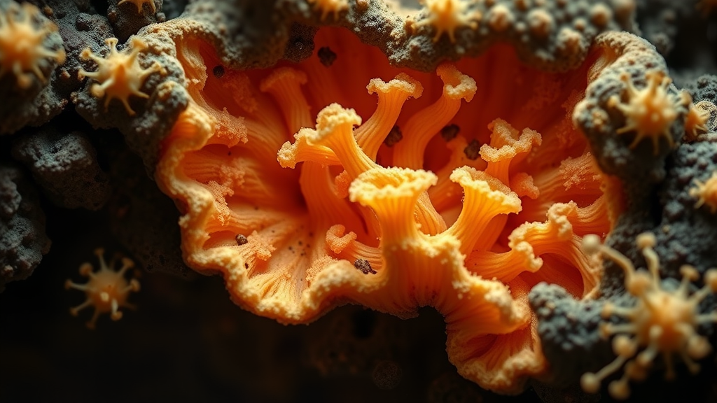
Chitin is to fungi what a load-bearing skeleton is to a building. Long chains of N-acetylglucosamine sugars are synthesized at the cell membrane and threaded outward, where they crystallize into microfibrils that reinforce the wall. Because human cells make no chitin at all, the enzymes responsible are theoretically ideal drug targets. Yet despite decades of interest, inhibitor development against chitin synthases has stalled, largely because researchers lacked a detailed picture of how these enzymes work, particularly the class II chitin synthases such as Chs1, which are essential for fungal viability. The new study changes that, presenting structures of CaChs1 at resolutions between 2.93 and 3.38 angstroms, sharp enough to trace individual amino acid side chains, bound substrates and even lipid molecules lodged inside the protein.
The technical achievement behind the structures is considerable. The team purified the enzyme from insect cells, verified its lipid cargo by liquid chromatography and mass spectrometry, and then froze thousands of single-particle specimens for imaging on cryo-electron microscopes. Computational classification of more than a hundred thousand micrographs allowed the researchers to sort the particles into distinct conformational states, effectively producing a molecular movie of the enzyme at work. They captured the enzyme in its resting, ligand-free form, in complexes with the sugar donor UDP-GlcNAc, and in a series of states showing the nascent chito-oligomer chain as it grows inside the catalytic site and begins its journey through the membrane-spanning translocation channel.
What emerges from these snapshots is a picture of a tightly choreographed machine. Chitin elongation and translocation are coupled to coordinated motion of the glycosyltransferase domain, the catalytic engine that adds sugar units one at a time, and the dimer interface, the region where two copies of the enzyme associate. As the growing polymer lengthens, the enzyme shifts between states that position the acceptor sugar for the next bond-forming step while simultaneously opening the channel through which the new chain is pushed toward the cell exterior. Molecular dynamics simulations carried out by collaborators at Lehigh University helped the team validate how the chitobiose product sits within the active site and how the polymer threads through the pore, complementing the static electron density maps with a dynamic view of the process.
The most striking discovery, however, concerns a small molecule called diynyl arylamine, abbreviated DA, a non-competitive inhibitor specific to CaChs1 that was previously known to block the enzyme without a clear mechanism. The structures show that DA does not bind at the catalytic site at all. Instead, it lodges within the chitin translocation channel itself, precisely where a regulatory phospholipid normally resides. By occupying this lipid-binding position, DA physically occludes the route through which the growing polymer must be extruded, jamming the machine in much the same way that a plug stops a pipe. This mode of inhibition, blocking product export rather than the chemical reaction itself, had been suspected for processive glycosyltransferases but had never been visualized in such atomic detail for a fungal chitin synthase.
The finding that a regulatory lipid sits inside the translocation channel adds an intriguing layer of biology. Lipids co-purifying with CaChs1 were identified as phosphatidylethanolamine species, and electron density attributable to a phospholipid was observed in the channel of the unliganded enzyme. The researchers propose that this lipid may help regulate the conformation of transmembrane helix 5 and an interfacial helix, elements that shift position when DA binds. In other words, the channel is not merely a passive tube but a gated, lipid-sensitive passage whose opening and closing is coordinated with the catalytic cycle. That a small drug can hijack this regulatory site suggests that channel-lipid interactions may be a general design principle among processive polymer-synthesizing enzymes, which include cellulose synthases in plants and hyaluronan synthases in vertebrates.
Perhaps the most clinically resonant result came from combination experiments. The team tested DA alongside nikkomycin Z, a nucleoside-derived natural product that inhibits class I chitin synthases by mimicking the sugar donor and occupying the catalytic site. Because the two compounds attack different parts of the chitin synthesis machinery, the researchers asked whether they would act synergistically. They did, and potently so. The combination showed strong synergy not only against C. albicans but also against Candida auris, the multidrug-resistant yeast that the World Health Organization has placed on its list of fungal priority pathogens. This matters because single-agent chitin synthase inhibitors have historically struggled: fungi can compensate for partial inhibition of one chitin synthase class by ramping up another. Hitting both the class I catalytic site and the class II translocation channel simultaneously appears to overwhelm that redundancy.
The work also fits into a broader renaissance in antifungal structural biology. In recent years, cryo-EM has illuminated the structures of chitin synthases from several organisms, including earlier studies of C. albicans class I enzymes and structures of fungal beta-1,3-glucan synthase bound to the frontline drug caspofungin, reported by an overlapping Duke team earlier in 2026. Together, these studies are mapping the two great polymer systems of the fungal wall, chitin and glucan, at atomic resolution. For the first time, medicinal chemists can see exactly where existing drugs bind, why some enzymes are resistant, and which pockets remain unexploited. The translocation channel of CaChs1 now joins that short list of validated, structure-guided targets.
Caution is warranted before declaring a new drug on the horizon. DA itself is a research tool rather than a medicine, and translating channel-blocking chemistry into a safe, potent antifungal will require optimization of selectivity, pharmacokinetics and formulation. The authors note that the DA-binding pocket is conserved among class II chitin synthases from Candida species and Saccharomyces cerevisiae, which is encouraging for spectrum but raises the question of how broadly across fungal pathogens such inhibitors could reach. Still, the conceptual advance is clear: the study establishes the chitin translocation channel as a druggable site and provides the structural foundation for rational design. At a time when antifungal drug resistance is rising and the clinical pipeline remains thin, every new angle of attack counts, and this one comes with an atomic-resolution map.
The structures and maps underlying the study have been deposited in public archives, including the Electron Microscopy Data Bank and the Protein Data Bank, so that laboratories worldwide can interrogate the channel, model their own inhibitor designs and test the lipid-regulation hypothesis. For a field that has waited more than four decades since chitin synthetase was first localized on the yeast plasma membrane for a molecular-level view of its target, the arrival of these structures marks a genuine turning point. The fungal wall’s most essential builder has finally been caught in the act, and its most vulnerable passage is now on the map.
Subject of Research: Cryo-EM structural analysis of the Candida albicans chitin synthase Chs1 and its druggable chitin translocation channel
Article Title: Cryo-EM structures of Candida albicans chitin synthase Chs1 reveal a druggable translocation channel
Article References: Cryo-EM structures of Candida albicans chitin synthase Chs1 reveal a druggable translocation channel. (n.d.). https://doi.org/10.1038/s41564-026-02485-2
Image Credits: AI Generated
DOI: 10.1038/s41564-026-02485-2
Keywords: Candida albicans, chitin synthase, cryo-EM, antifungal resistance, translocation channel, diynyl arylamine, nikkomycin Z, Candida auris, fungal cell wall, drug discovery, structural biology, glycosyltransferase
Cite Scienmag News
Jason Bradley. (September 24, 2026). Fungal cell wall machine exposed: chitin synthase channel offers new antifungal target. Scienmag. https://scienmag.com/fungal-cell-wall-machine-exposed-chitin-synthase-channel-offers-new-antifungal-target/
Jason Bradley. "Fungal cell wall machine exposed: chitin synthase channel offers new antifungal target." Scienmag, 24 September 2026, https://scienmag.com/fungal-cell-wall-machine-exposed-chitin-synthase-channel-offers-new-antifungal-target/. Accessed 5 October 2026.
Jason Bradley. "Fungal cell wall machine exposed: chitin synthase channel offers new antifungal target." Scienmag. September 24, 2026. https://scienmag.com/fungal-cell-wall-machine-exposed-chitin-synthase-channel-offers-new-antifungal-target/


