


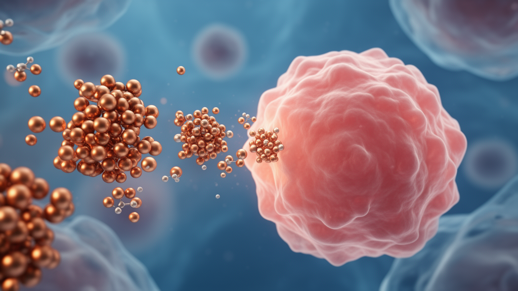
Copper has quietly become one of the most intriguing materials in modern medicine. A comprehensive review published in Frontiers in Nanotechnology by researchers at Chapman University School of Pharmacy and Marshall B. Ketchum University synthesizes the rapidly expanding field of copper nanoparticles (CuNPs), arguing that these tiny metallic particles could reshape how drugs and genes are delivered to tumors while simultaneously killing cancer cells through mechanisms that conventional chemotherapy cannot touch. The review, led by Keykavous Parang and colleagues, integrates the chemistry, biological fate, and therapeutic applications of CuNPs into a single framework, linking the recently discovered cell death pathway known as cuproptosis to the rational design of next-generation nanomedicine platforms.
What makes copper nanoparticles so attractive is a combination of properties that few other materials can match. Their large surface area allows efficient drug loading, their size can be tuned during synthesis, their surfaces can be easily modified with polymers, peptides, antibodies, and targeting ligands, and their production cost is dramatically lower than that of gold or silver alternatives. Beyond serving as passive carriers, CuNPs possess intrinsic antimicrobial, anticancer, anti-inflammatory, and photothermal activities, meaning the delivery vehicle itself contributes to the therapeutic effect. The particles can be fabricated as metallic copper, cuprous oxide, or cupric oxide, each phase exhibiting distinct reactivity, optical behavior, and biological interactions that researchers can exploit for different applications.
The synthesis route fundamentally determines what a copper nanoparticle can do. Wet-chemical reduction, the most widely used approach, employs copper salts and reducing agents such as ascorbic acid or sodium borohydride to yield zero-valent nanoparticles, with capping agents preventing aggregation and oxidation. Green synthesis has emerged as a critical alternative, using plant extracts rich in polyphenols, flavonoids, and alkaloids that simultaneously reduce copper ions and stabilize the resulting nanostructures. By carefully controlling pH, temperature, extract concentration, and reaction time, researchers can produce spherical, rod-like, or flower-shaped particles typically ranging from 5 to 50 nanometers. Surface engineering with polyethylene glycol, polyvinylpyrrolidone, silica shells, or organic ligands then stabilizes the particles in physiological environments, a crucial step for any biomedical deployment.
Once inside the body, copper nanoparticles follow a well-defined biological journey. After administration via inhalation, oral, dermal, or injection routes, they enter systemic circulation, acquire a protein corona, and preferentially accumulate in the liver and spleen. At the cellular level, lysosomal dissolution releases copper ions that integrate into the body’s endogenous copper homeostasis pathways. Toxicity arises through oxidative stress, mitochondrial dysfunction, membrane damage, inflammation, and genotoxicity. Elimination occurs predominantly through hepatobiliary excretion, with a secondary renal route. Animal studies have revealed a clear inverse size-dependent toxicity profile: oral lethal dose values ranged from 1,022 mg/kg for 30-nanometer particles to over 5,000 mg/kg for one-micrometer copper microparticles, with smaller particles showing greater absorption efficiency due to their increased surface-area-to-volume ratio.
The anticancer mechanisms of CuNPs are particularly compelling. Upon tumor targeting and cellular uptake, intracellular release of copper ions drives the generation of reactive oxygen species, culminating in oxidative DNA damage, activation of apoptotic pathways, and induction of cuproptosis, a copper-dependent cell death mechanism distinct from classical apoptosis. Cancer cells are especially vulnerable to copper-mediated oxidative damage because they already operate at elevated basal reactive oxygen species levels with reduced antioxidant capacity. The discovery of cuproptosis by Tsvetkov and colleagues demonstrated that excess copper directly binds to lipoylated proteins of the tricarboxylic acid cycle, causing their aggregation and the loss of iron-sulfur cluster proteins, ultimately inducing proteotoxic stress. The enzyme ferredoxin-1 was identified as a central upstream regulator, reducing cupric copper to the more toxic cuprous form. Because cuproptosis depends on mitochondrial metabolism, cancer cells relying heavily on oxidative phosphorylation are especially susceptible, opening a selective therapeutic window.
Preclinical results from specific nanoparticle formulations illustrate the therapeutic potential in striking detail. Researchers developed copper diethyldithiocarbamate nanoparticles using a stabilized metal ion ligand complex method that achieved near 100 percent drug-loading efficiency, representing a greater than 200-fold improvement over traditional micelle formulations. These sub-100-nanometer particles demonstrated potent activity against paclitaxel-resistant prostate cancer cells with half-maximal inhibitory concentrations as low as 85 nanomolar, while also bypassing P-glycoprotein-mediated drug efflux, a major mechanism of multidrug resistance. In xenograft models, intravenous administration significantly reduced tumor volume without notable body weight loss or systemic toxicity. Separately, lactoferrin-coated copper nanoparticles showed strong synergistic activity against malignant melanoma cells, with combination index values indicating powerful cooperativity between copper and the milk protein carrier.
Beyond direct cytotoxicity, copper nanoparticles excel as controlled and targeted drug delivery vehicles. The enhanced permeability and retention effect allows nanoparticles to passively accumulate in tumor tissue by exploiting the leaky vasculature characteristic of solid tumors. Active targeting can be achieved by decorating particle surfaces with folic acid, antibodies, aptamers, or peptides that bind receptors overexpressed on cancer cells. Stimuli-responsive platforms exploit the tumor microenvironment’s distinctive biochemistry, including acidity, elevated glutathione, hypoxia, and excessive reactive oxygen species, to release drugs specifically within tumor tissue while remaining inert in healthy tissue. Copper-based nanoplatforms are particularly suited to pH-responsive delivery because copper oxides readily dissolve under the mildly acidic conditions of endosomes and lysosomes, triggering controlled ion and drug release precisely where needed.
The versatility extends to gene delivery, where the positive surface charge of functionalized copper nanoparticles enables electrostatic complexation with negatively charged nucleic acids. Researchers demonstrated that plant-derived copper oxide nanoparticles functionalized with chitosan and polyethylene glycol, then conjugated to folate, successfully bound and protected a reporter plasmid, enabling targeted gene delivery to folate receptor-overexpressing cervical cancer cells. More advanced bimetallic drug-gene co-delivery systems utilize copper redox cycling to inhibit the glutathione axis while simultaneously delivering small interfering RNA targeting resistance pathways, activating immunogenic ferroptosis and amplifying antitumor immunity. Photothermal and photodynamic therapy represent additional modalities, with copper sulfide nanoparticles converting near-infrared light into localized heat that destroys tumor cells while minimizing damage to surrounding healthy tissue.
Despite this remarkable promise, the field faces substantial translational barriers. The same redox activity that underlies anticancer potency also generates the principal safety liability, as uncontrolled copper ion release in non-target tissues induces oxidative stress, membrane damage, and genotoxicity in healthy cells. The overwhelming majority of published studies remain in vitro, with essentially no clinical trial data for copper nanoparticle therapeutics as injectable medicines. Manufacturing scalability, batch-to-batch reproducibility, long-term physicochemical stability, and the absence of harmonized regulatory frameworks for copper-based nanomedicines collectively represent significant obstacles. The review’s authors argue that investment in rigorous in vivo pharmacokinetic and toxicology studies, standardized characterization protocols, and well-powered animal efficacy studies in immunocompetent models will determine whether copper nanoparticles fulfill their considerable promise as next-generation cancer nanomedicines.
The convergence of cuproptosis-based immunotherapy with copper nanoparticle platforms represents what the researchers consider a transformative opportunity. Recent preclinical work demonstrates that copper-mediated cell death synergizes powerfully with immune checkpoint blockade by inducing immunogenic cell death and remodeling immunosuppressive tumor microenvironments. Future designs are expected to increasingly incorporate biomimetic coatings, cell membrane camouflage, and artificial intelligence-assisted nanoparticle optimization to sharpen tumor specificity and reduce systemic exposure. As the field stands at a critical inflection point, the extraordinary potential of copper nanoparticles in drug delivery and anticancer therapy is matched only by the extraordinary complexity of the engineering, toxicological, and regulatory challenges that must be solved before these versatile particles can deliver tangible benefit to patients.
Subject of Research: Copper nanoparticles for drug delivery and anticancer therapy
Article Title: Copper nanoparticles and their future in drug delivery and anticancer therapy
Article References: Parang, K., Vadlapatla, R., Charles, C., Tang, K., Nguyen, T., Shirazi, A. N., & Koomer, A. (2026). Copper nanoparticles and their future in drug delivery and anticancer therapy. Frontiers in Nanotechnology, 8, Article 1906191. https://doi.org/10.3389/fnano.2026.1906191
Image Credits: AI Generated
DOI: 10.3389/fnano.2026.1906191
Keywords: copper nanoparticles, drug delivery, cancer therapy, cuproptosis, nanomedicine, photothermal therapy, gene delivery, targeted therapy, Copper, nanoparticles, future, drug
Cite Scienmag News
Nathaniel Bowman. (October 16, 2026). Copper Nanoparticles Emerge as Multifunctional Weapons in Drug Delivery and Cancer Therapy. Scienmag. https://scienmag.com/copper-nanoparticles-emerge-as-multifunctional-weapons-in-drug-delivery-and-cancer-therapy/
Nathaniel Bowman. "Copper Nanoparticles Emerge as Multifunctional Weapons in Drug Delivery and Cancer Therapy." Scienmag, 16 October 2026, https://scienmag.com/copper-nanoparticles-emerge-as-multifunctional-weapons-in-drug-delivery-and-cancer-therapy/. Accessed 16 October 2026.
Nathaniel Bowman. "Copper Nanoparticles Emerge as Multifunctional Weapons in Drug Delivery and Cancer Therapy." Scienmag. October 16, 2026. https://scienmag.com/copper-nanoparticles-emerge-as-multifunctional-weapons-in-drug-delivery-and-cancer-therapy/


