


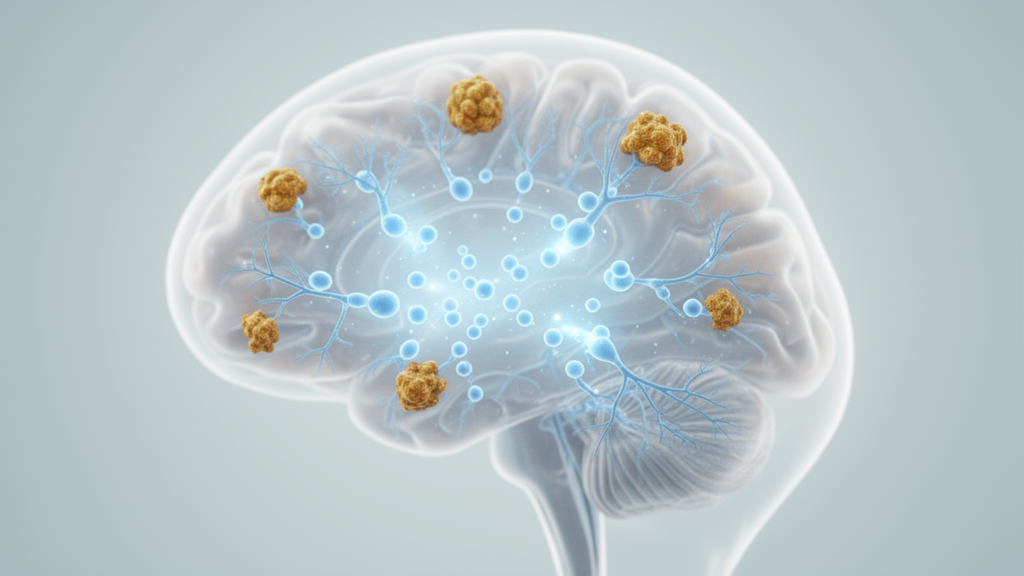
Chronic treatment with neuropeptide S, a small wakefulness-promoting brain signaling molecule, has been shown to improve disturbed sleep architecture and reduce amyloid-related brain pathology in a mouse model of Alzheimer’s disease. The finding, reported in Frontiers in Neurology by a team at Jiangnan University in China, is notable because it runs counter to what the same molecule does when given acutely: a single dose of neuropeptide S (NPS) is known to keep rodents awake. When delivered daily into the brain ventricles for two weeks, however, NPS increased non-rapid eye movement (NREM) sleep, reduced wakefulness, and was accompanied by lower levels of amyloid precursor protein, its phosphorylated form, and amyloid-β plaque burden in APPswe/PS1dE9 (APP/PS1) mice, a widely used double-transgenic model of Alzheimer’s disease.
Sleep disturbances are among the most common and earliest non-cognitive symptoms of Alzheimer’s disease. Patients experience fragmented nighttime sleep, daytime sleepiness, and disrupted 24-hour rest–activity rhythms, and similar abnormalities have been documented in transgenic mouse models. People with amnestic mild cognitive impairment, a condition that often precedes Alzheimer’s disease, already show measurable changes during sleep, including fewer stage-2 sleep spindles and less time in deep NREM sleep. Observational studies have linked poorer sleep quality to amyloid deposition even in cognitively unimpaired adults, and longitudinal evidence associates insomnia symptoms and sleep-disordered breathing with increased risk of later cognitive impairment and dementia. These observations underpin a proposed bidirectional relationship in which sleep disruption and amyloid pathology reinforce one another.
NPS is a conserved 20-amino-acid neuropeptide produced mainly in the peri-locus coeruleus and parabrachial nucleus of the brainstem. It binds the NPS receptor (NPSR), a Gαq-coupled receptor distributed widely across the brain, with high density in the amygdala, thalamus, and prefrontal cortex. The NPS–NPSR system is known to enhance cognition by facilitating hippocampal long-term potentiation, to reduce anxiety, and to regulate the sleep–wake cycle. Acute intracerebroventricular administration of NPS increases wakefulness and suppresses both NREM and REM sleep in rodents, an effect accompanied by activation of histaminergic and orexinergic neurons in the posterior hypothalamus. What remained unclear was how chronic NPS exposure affects sleep in a disease context, where sleep regulation itself is pathological.
The researchers began by characterizing the NPSR system in the disease model. Western blotting and immunohistochemistry revealed lower NPSR protein levels and fewer NPSR-positive cells in both the hypothalamus and the hippocampus of APP/PS1 mice compared with wild-type littermates. Because the measurements were taken at a single age, the authors note that this reduction cannot be shown to precede or contribute causally to the sleep abnormalities, but it establishes that the receptor system is altered in regions central to both sleep regulation and memory.
Sleep–wake architecture was then compared between saline-treated wild-type and APP/PS1 mice, all 8-month-old males, using continuous polysomnographic recording over one complete light–dark cycle. The APP/PS1 mice spent more time awake and less time in NREM sleep during the light phase and over 24 hours, and less time in REM sleep during the light phase and over 24 hours. They also had more wake episodes and fewer REM episodes during the light phase, more NREM episodes during the dark phase, and shorter mean NREM episodes in both phases — a fragmented sleep pattern that mirrors clinical descriptions of Alzheimer’s-related sleep disruption.
After 14 days of daily intracerebroventricular NPS at 1 nmol per day, the picture changed. Compared with saline-treated APP/PS1 mice, the NPS-treated animals showed reduced wakefulness and increased NREM sleep, with the largest differences during the light phase and over 24 hours. REM sleep time did not differ significantly between the two groups. NPS-treated mice had fewer wake episodes during the light phase, fewer NREM episodes in both phases, and fewer REM episodes during the dark phase, while mean NREM episode duration lengthened in both phases. In other words, sleep became not only more abundant but also more consolidated. The authors emphasize that this chronic response differs from the acute wake-promoting effect of NPS, but because treatment duration and disease background were not varied independently in the study, the reason for the reversal remains uncertain. The absence of a wild-type group receiving NPS also means it cannot be determined whether the sleep response is specific to the disease model.
The amyloid findings paralleled the sleep changes. Saline-treated APP/PS1 mice had higher levels of amyloid precursor protein (APP) and APP phosphorylated at threonine 668 in both the hypothalamus and hippocampus than wild-type controls. After NPS treatment, both APP and p-APP levels fell in both regions, and the percentage of 4G8-immunoreactive amyloid-β area in the hippocampus was reduced. Co-staining for Iba1, a microglial marker, and 4G8 showed microglia in close proximity to plaques in both treated and untreated groups, but the mean number of 4G8-positive plaques per field was significantly lower after NPS treatment. The authors caution that these histological measures do not directly quantify microglial uptake of amyloid-β, and that lower APP and p-APP levels leave open the possibility of reduced amyloid production rather than enhanced clearance. Sleep itself may contribute, since prior work shows sleep facilitates interstitial solute and amyloid-β clearance in the brain, but the respective roles of these processes were not tested.
To probe whether amyloid can drive sleep changes in the treated animals, the team gave NPS-pretreated APP/PS1 mice a single intracerebroventricular injection of synthetic Aβ₁–₄₂ or a matched saline injection 24 hours before recording. The Aβ-challenged mice spent more time awake and less time in NREM sleep than NPS-treated mice receiving saline, mainly during the dark phase and over 24 hours, and their NREM episodes were shorter during the light phase. The authors point out important caveats: this pattern only partly overlaps with the effects of chronic NPS, which were most evident in the light phase, and without a comparison group receiving Aβ₁–₄₂ without prior NPS treatment it is unclear whether the response depended on NPS pretreatment. The Aβ preparation also contained dimethyl sulfoxide, whereas the comparison injection was saline alone, so vehicle-related effects cannot be excluded.
Parallel experiments in BV2 cells, an immortalized microglial cell line, explored a candidate cellular mechanism. When cells were exposed to Aβ₁–₄₂ together with NPS, cell-associated amyloid-β levels rose significantly after one hour of co-treatment, though not at 0.5, 1.5, or 2 hours, and more Iba1/4G8 double-positive cells were counted. The authors note these measurements cannot distinguish increased uptake from reduced degradation, surface-bound from internalized amyloid, or differences in cell density. In a washout protocol, cell-associated amyloid levels were higher with NPS co-treatment before washout but similar between the two Aβ-exposed groups 24 hours after extracellular amyloid was removed — consistent with turnover but not proof of accelerated degradation. After 24 hours of treatment, the NPS co-treatment group showed higher LC3B-II and lower p62 levels, markers consistent with autophagy-related changes, though single-endpoint measurements without lysosomal inhibition cannot establish increased autophagic flux. NPS co-treatment also increased the ratio of phosphorylated to total ERK, while p38, JNK, and AKT phosphorylation ratios were unchanged, and the ERK inhibitor SCH772984 attenuated both the autophagy-marker changes and the cell-associated amyloid-β changes, supporting possible ERK involvement.
The authors are careful about how far these cellular findings carry. The autophagy-marker and amyloid experiments used different NPS concentrations, 300 nanomolar and 100 nanomolar respectively, so they do not directly establish an ERK–autophagy–amyloid degradation sequence under the same conditions, and whether the BV2 responses contribute to the effects seen in living mice remains unresolved. Further limitations include the restriction of the animal work to 8-month-old males, the use of an immortalized cell line rather than primary microglia, histology based on selected coronal sections and fields, and the fact that Iba1 staining alone does not establish a microglial activation phenotype. Within those bounds, the study demonstrates that chronic central NPS administration improved selected aspects of sleep architecture and was associated with reduced amyloid-related pathology in APP/PS1 mice, while pointing to ERK signaling and autophagy-related processes as candidate mechanisms for how the neuropeptide system might influence cellular amyloid handling. Whether the same approach could translate to patients with Alzheimer’s-related sleep disturbance is a question the mouse and cell data, on their own, cannot answer.
Subject of Research: Effects of chronic neuropeptide S treatment on sleep architecture and amyloid-β pathology in an Alzheimer's disease mouse model
Article Title: Chronic neuropeptide S treatment improves sleep architecture and reduces Aβ-related pathology in APP/PS1 mice
Article References: Zhao, Y., Liu, M., Zhou, Y., Cui, M., Cai, N., Wang, H., Liu, G., Sun, H., Kong, X., & Zhao, P. (2026). Chronic neuropeptide S treatment improves sleep architecture and reduces Aβ-related pathology in APP/PS1 mice. Frontiers in Neurology, 17, Article 1825666. https://doi.org/10.3389/fneur.2026.1825666
Image Credits: AI Generated
DOI: 10.3389/fneur.2026.1825666
Keywords: Alzheimer's disease, neuropeptide S, NPSR, sleep architecture, amyloid-β, APP/PS1 mice, NREM sleep, microglia, BV2 cells, ERK signaling, autophagy, polysomnography
Cite Scienmag News
Cassandra Pierce. (October 26, 2026). Chronic Neuropeptide S Treatment Restores Sleep and Cuts Amyloid Pathology in Alzheimer’s Mice. Scienmag. https://scienmag.com/chronic-neuropeptide-s-treatment-restores-sleep-and-cuts-amyloid-pathology-in-alzheimers-mice/
Cassandra Pierce. "Chronic Neuropeptide S Treatment Restores Sleep and Cuts Amyloid Pathology in Alzheimer’s Mice." Scienmag, 26 October 2026, https://scienmag.com/chronic-neuropeptide-s-treatment-restores-sleep-and-cuts-amyloid-pathology-in-alzheimers-mice/. Accessed 26 October 2026.
Cassandra Pierce. "Chronic Neuropeptide S Treatment Restores Sleep and Cuts Amyloid Pathology in Alzheimer’s Mice." Scienmag. October 26, 2026. https://scienmag.com/chronic-neuropeptide-s-treatment-restores-sleep-and-cuts-amyloid-pathology-in-alzheimers-mice/


