


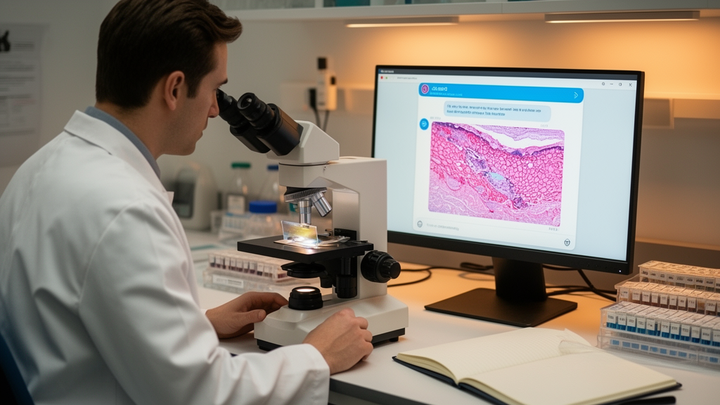
A team of dermatologists at the University of California, Los Angeles has put two of the world’s most widely used artificial intelligence chatbots to an unusual test: instead of asking them to analyze photographs of skin lesions, the researchers showed them histologic images, the stained tissue sections that pathologists examine under the microscope when making a definitive cancer diagnosis. The study, published as a research letter in the Archives of Dermatological Research, evaluated how accurately ChatGPT and Gemini could identify basal cell carcinoma, the most common form of skin cancer in humans, from microscopic images alone.
The distinction between clinical and histologic evaluation matters because basal cell carcinoma is ultimately confirmed by tissue examination rather than by visual inspection alone. When a dermatologist suspects a lesion clinically, a biopsy is taken and processed into thin, stained sections—most commonly with hematoxylin and eosin—that reveal the cellular architecture of the tumor. Pathologists then look for characteristic features such as nests of basaloid cells, peripheral palisading, and stromal retraction artifacts. This means that any tool hoping to assist in diagnosis at the definitive stage must operate in the domain of microscopy, not dermatoscopy, which is precisely the niche the UCLA team chose to probe.
Basal cell carcinoma accounts for the overwhelming majority of non-melanoma skin cancers and, despite its low metastatic potential, can cause significant local tissue destruction when diagnosis is delayed. Its histologic presentation, however, is not uniform. Subtypes such as nodular, superficial, infiltrative, and morpheaform variants each carry distinct microscopic patterns, and some can be confused with benign adnexal tumors or other cutaneous malignancies. This heterogeneity is one reason automated classification of basal cell carcinoma has historically been challenging, even for purpose-built deep learning systems trained on large annotated slide datasets.
The choice of ChatGPT and Gemini as test subjects reflects a broader shift in how medical researchers evaluate artificial intelligence. Earlier generations of diagnostic algorithms were narrow models trained specifically on medical images, often requiring curated datasets and supervised fine-tuning. Large multimodal language models, by contrast, are general-purpose systems that can accept images and text prompts without task-specific training. Their apparent versatility has fueled speculation that they might serve as accessible diagnostic aids, particularly in settings where specialist expertise is scarce. But general-purpose training also means these models were never explicitly optimized for histopathology, and their internal reasoning about tissue morphology remains largely opaque to researchers.
The UCLA group’s work builds directly on their own earlier investigation, in which the same team evaluated ChatGPT and Gemini using clinical photographs of non-melanoma skin cancer rather than microscopic images. That prior study, published in the same journal, framed a question that has become increasingly common in dermatology informatics: whether conversational AI systems accessible to ordinary patients could function as preliminary screening tools. By extending the analysis to histologic images, the researchers effectively moved one step deeper into the diagnostic pipeline, testing whether the models perform better or worse when given the kind of information a pathologist would actually use to render a definitive diagnosis.
Their study also sits within a growing body of literature on machine learning and basal cell carcinoma specifically. A 2023 analysis published in the journal Pathology examined artificial intelligence systems for diagnosing basal cell carcinoma and distinguishing it from histologic mimics, underscoring that even dedicated image-classification models must contend with look-alike lesions that challenge human experts. On the melanoma side, a widely cited 2019 study demonstrated that a deep learning system could outperform a panel of eleven pathologists in classifying histopathological melanoma images, a result that energized the field but also highlighted how much curated training data such systems require. The contrast between those bespoke systems and today’s general-purpose chatbots is central to understanding what the new research letter does and does not demonstrate.
Another relevant precedent is a 2025 study published in JMIR Dermatology that assessed ChatGPT-4’s ability to identify diverse skin lesions against squamous cell carcinoma and basal cell carcinoma. That work, like the UCLA investigation, focused on commercially available models rather than custom-built classifiers, and it reflects a methodological trend: researchers are increasingly treating frontier AI models as black boxes whose diagnostic competence can be measured empirically, without access to their training data or internal architecture. This approach has practical appeal but also limits the interpretability of results, since researchers cannot easily determine why a model succeeds on one image and fails on another.
The images used in the UCLA analysis were obtained with permission from Pathology Outlines, a publicly accessible pathology reference database widely used by trainees and practicing pathologists. This sourcing choice has both advantages and caveats worth considering. On the positive side, using publicly available images makes the study reproducible: any interested reader can view the same material the models were shown. It also avoids the logistical and ethical burdens of assembling a new biopsy archive. On the other hand, images published in a reference database are often chosen precisely because they are classic, well-demonstrated examples of a disease, which may make them easier to classify than the ambiguous, poorly oriented, or artifact-laden sections that populate real-world surgical pathology workflows. Performance on curated teaching images may therefore represent an optimistic ceiling rather than a realistic floor.
It is also worth noting that histologic image interpretation involves contextual information that a static image may not fully convey. Pathologists routinely integrate clinical data—patient age, lesion location, prior history, and the gross appearance of the biopsy—into their diagnostic reasoning. A model shown only a cropped microscopic field lacks this context, as does, in fairness, any reader of the same image. Whether the researchers provided clinical vignettes alongside the images, and how the models’ outputs were scored against a reference standard, are details that shape how the accuracy figures should be interpreted. Research letters, by their brevity, often leave such methodological questions for readers to weigh.
The publication timeline of the study offers its own context. The manuscript was received in May 2026, revised in June, and accepted in August, with publication on the first of September that year in volume 318 of the journal. The rapid turnaround is typical of research letters, which are designed to communicate concise findings quickly rather than to present exhaustive analyses. The authors declared no competing interests and reported no external funding, which suggests the work was conducted as an academic side project rather than an industry-sponsored evaluation—a common pattern in this literature, and one that underscores both the accessibility of the methods and the modest scale typical of such studies.
The authorship composition is also informative. The team spans the Division of Dermatology at UCLA and the David Geffen School of Medicine, combining clinicians with a medical student co-author. The division of labor described in the paper—conceptualization and study design by two senior authors, data collection by one, and analysis and manuscript preparation by another—reflects the increasingly common structure of AI-evaluation studies in academic dermatology, where clinical expertise guides the framing while trainees often carry out the hands-on testing and writing.
For clinicians and patients alike, the practical takeaway from this line of research is one of cautious interest rather than immediate application. Even if ChatGPT and Gemini perform respectably on curated histologic images, no regulatory framework currently permits their use as diagnostic devices, and their outputs can vary between queries in ways that are poorly understood. The models’ training data are not disclosed in detail, raising the possibility that images similar to those in public pathology databases were encountered during training, which would inflate apparent accuracy. Independent validation on fresh, institution-specific biopsy material remains the gold standard for establishing whether general-purpose AI can meaningfully support histopathologic diagnosis.
Nevertheless, the trajectory of this research area is notable. Within just a few years, the question has shifted from whether artificial intelligence can classify skin cancer at all—largely settled by dedicated deep learning systems—to whether widely available conversational models can do so without any specialized training. The UCLA team’s paired studies, moving from clinical photographs to histologic sections, map the diagnostic pipeline itself and ask where in that pipeline general-purpose AI might prove useful. As multimodal models continue to improve and as larger, more diverse validation datasets become available, studies of this kind will serve as useful benchmarks, charting both the progress and the persistent limitations of AI in the microscopic diagnosis of the world’s most common skin cancer.
Subject of Research: Diagnostic accuracy of large language model chatbots in identifying basal cell carcinoma from histologic images
Article Title: Evaluation of diagnostic accuracy of ChatGPT and gemini in identification of basal cell carcinoma in histologic images
Article References: Khan, S., Hendrie, A., Modiri, O., & Davis, J. (2026). Evaluation of diagnostic accuracy of ChatGPT and gemini in identification of basal cell carcinoma in histologic images. Archives of Dermatological Research, 318(1), Article 388. https://doi.org/10.1007/s00403-026-04836-9
Image Credits: AI Generated
DOI: 10.1007/s00403-026-04836-9
Keywords: artificial intelligence, ChatGPT, Gemini, basal cell carcinoma, dermatopathology, histopathology, large language models, diagnostic accuracy, skin cancer, machine learning, medical imaging, pathology
Cite Scienmag News
Ophelia Keating. (October 16, 2026). ChatGPT and Gemini Face a Histology Test: How Well Can AI Chatbots Spot Basal Cell Carcinoma Under the Microscope? Scienmag. https://scienmag.com/chatgpt-and-gemini-face-a-histology-test-how-well-can-ai-chatbots-spot-basal-cell-carcinoma-under-the-microscope/
Ophelia Keating. "ChatGPT and Gemini Face a Histology Test: How Well Can AI Chatbots Spot Basal Cell Carcinoma Under the Microscope?" Scienmag, 16 October 2026, https://scienmag.com/chatgpt-and-gemini-face-a-histology-test-how-well-can-ai-chatbots-spot-basal-cell-carcinoma-under-the-microscope/. Accessed 16 October 2026.
Ophelia Keating. "ChatGPT and Gemini Face a Histology Test: How Well Can AI Chatbots Spot Basal Cell Carcinoma Under the Microscope?" Scienmag. October 16, 2026. https://scienmag.com/chatgpt-and-gemini-face-a-histology-test-how-well-can-ai-chatbots-spot-basal-cell-carcinoma-under-the-microscope/


