


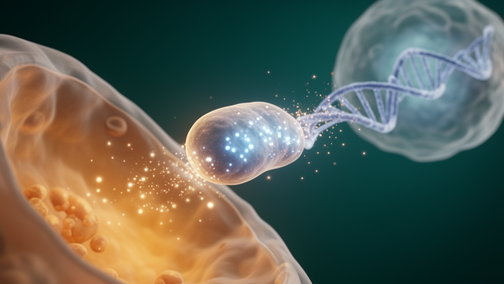
Every living cell emits an almost unimaginably faint shimmer of light. This ultra-weak photon emission, or UPE, has been measured for decades in the ultraviolet, visible, and near-infrared ranges, yet most biologists have treated it as metabolic exhaust—nothing more than the byproduct of reactive molecules colliding inside the cell. A new review and position paper published in Frontiers in Photobiology argues that this dismissal may be a profound mistake. Roeland Van Wijk and Eduard P. A. Van Wijk of Meluna Research in the Netherlands weave together two historically separate strands of biophoton research into a single, provocative hypothesis: the light produced by mitochondria may be absorbed by nuclear chromatin, stored there, and re-emitted in delayed pulses that feed back onto the cell’s energy machinery, creating a photonic dialogue between the cell’s power plants and its genome.
The story begins nearly a century ago. In the 1920s, the Russian biologist Alexander Gurwitsch performed an experiment that remains one of the strangest in biology: he showed that light emitted from the tip of one onion root could stimulate cell division in a second root placed nearby, even when all known chemical signals were excluded. Gurwitsch called this “mitogenetic radiation” and introduced the idea of a biological “field” of forces governing cellular behavior. By the 1930s he had discovered that atmospheric oxygen was required for the effect, hinting that the radiation arose from reactions involving short-lived free radicals—species whose chemistry was only beginning to be understood.
Modern biophysics has largely vindicated the physical basis of those early observations. When single electrons leak from the mitochondrial electron transport chain onto molecular oxygen, they form superoxide anions, which are converted to hydrogen peroxide and, ultimately, to singlet oxygen—an electronically excited state of O2. Singlet oxygen emits photons at roughly 780 nanometers, and its dimers produce characteristic bands near 585, 635, and 703 nanometers. Excited carbonyl compounds generated during the peroxidation of mitochondrial membrane lipids add emissions in the 500 to 560 nanometer range, while some radical recombination reactions release enough energy to produce ultraviolet photons between 230 and 300 nanometers. In other words, the entire spectrum from ultraviolet to infrared can be traced back to superoxide formation and lipid peroxidation, with early studies attributing up to 75 percent of visible emission to free radical recombination.
What has changed in recent years is the recognition that this photon production is not a steady drip but a pulsatile, coordinated phenomenon. Beginning in the early 2000s, researchers using targeted fluorescent probes discovered “mitochondrial superoxide flashes”—transient bursts of superoxide generation within the mitochondrial matrix lasting roughly 10 to 15 seconds. Subsequent imaging showed that calcium uptake, membrane potential, and pH all fluctuate in synchrony during individual flashes, indicating that each event reflects a coordinated, transient reorganization of mitochondrial physiology. In skeletal muscle and cardiac tissue, these flashes often occur simultaneously among spatially clustered mitochondria. The prevailing picture has therefore shifted from mitochondria as isolated, passive producers of ATP and reactive oxygen species to mitochondria as components of an interconnected network whose output is regulated through amplitude and frequency modulation—much like AM and FM radio signals.
Here the arithmetic becomes striking. A photomultiplier tube placed above a liver cell culture containing about six million cells typically records no more than a few hundred photons per second per square centimeter—roughly one detected photon per second per 2,000 cells. But estimates based on cellular oxygen consumption tell a very different story. An average rat cell processes on the order of 10^12 oxygen molecules per day, and if about 2 percent undergo single-electron reduction to superoxide, with at least 1 percent of those reactions contributing to photon production, each cell generates roughly 2,300 photons per second. The discrepancy between calculated production and detected emission implies that intracellular absorption of mitochondrially produced light must be extraordinarily high—on the order of a millionfold or greater. The question then becomes: what inside the cell is soaking up all this light?
The authors argue that the prime candidate is the nucleus, and specifically chromatin—the complex of DNA and associated proteins. The nucleus occupies about 10 percent of cell volume and is surrounded by thousands of mitochondria in the perinuclear cytoplasm, so synchronously emitted photon pulses are likely to impinge upon it with high probability. Supporting evidence comes from decades of delayed luminescence experiments, in which cells are exposed to external light and then monitored in complete darkness as they re-emit stored photons. Cell fractionation studies showed that the cytoplasm, including mitochondria and ribosomes, exhibits little long-term photon storage, while intact isolated nuclei retain even more capacity than whole cells. Crucially, when DNA was separated from its associated proteins, neither fraction alone retained significant luminescence—photon storage is an emergent property of chromatin as an integrated structure, not of naked DNA.
Even more intriguing, photon storage capacity varies systematically with cellular differentiation. In classic experiments spanning 17 mammalian cell lines, poorly differentiated tumor cells emitted photons rapidly and intensely, indicating minimal storage, while normal, well-differentiated cells showed slow, prolonged emission consistent with high storage capacity. Well-differentiated hepatoma cells fell in between. Normal liver cells even retained more photons at higher culture densities, consistent with reabsorbing light emitted by their neighbors, whereas poorly differentiated tumor cells continued to pour out photons regardless of density. Whether this optical signature reflects cause or consequence of the differentiated state remains open, but the correlation is consistent across tissues and species.
The paper then confronts a fundamental question that chromatin biology has largely left unanswered: what physical principle drives chromatin motion at the molecular level? Contemporary models depict the genome as a dynamic, fluid mosaic—compacted a thousandfold yet constantly remodeled by ATP-dependent complexes, transcription bubbles traveling along genes, and topoisomerases resolving topological stress. The authors propose that light provides a key part of the answer through excitonic processes: delocalized electron–hole excitations that can propagate along the regular hydrogen-bonded geometry of protein alpha-helices and through stacked DNA bases, persisting for up to milliseconds before relaxing with red-shifted photon emission. Recent ultrafast spectroscopy has shown that exciton lifetimes in DNA depend on base sequence, with guanine-rich tracts localizing charge and adenine-rich tracts facilitating migration. Charge delocalization within chromatin could transiently alter local conformation and electrostatic potential, potentially modulating the affinity of regulatory proteins for DNA.
Putting all the pieces together, the authors advance a speculative but testable model: two alternating, pulsatile photonic spectra—one originating from mitochondrial emission, the other from delayed chromatin–DNA luminescence—together sustain a dynamic equilibrium between mitochondrial energy production and the energy demands of chromatin organization and gene regulation. Mitochondrial light would act as a primary trigger exciting chromatin, while chromatin’s red-shifted delayed emission would feed back onto the mitochondrial network, tuning respiration to the genome’s activity. Notably, the action spectrum of low-intensity light therapy, which stimulates cytochrome c oxidase and ATP synthesis, does not match the primary emission bands of mitochondrial reactive oxygen species—but it could match red-shifted, delayed luminescence from chromatin, offering a possible resolution of the long-standing paradox by which light therapy transiently increases reactive oxygen species yet protects tissue from oxidative damage.
The authors are careful to frame this as a hypothesis in need of experimental verification, and they outline a concrete research program: characterizing spontaneous emission spectra under physiological and stress conditions using high-sensitivity multi-band filter systems, using the 5-to-10-second rhythm of mitochondrial pulsations to deposit light energy into chromatin with narrowly defined wavelengths, and recording wavelength-specific delayed emission with modern time-resolved photomultiplier equipment. Recent work showing that red and near-infrared light treatment alters biophoton emission in stressed but not healthy cell cultures offers an early encouraging sign. If the model survives such scrutiny, biophotons would graduate from metabolic noise to a genuine regulatory layer—one in which the cell’s inner light helps choreograph the dance of its own genome.
Subject of Research: The proposed regulatory role of ultra-weak photon emission in mitochondrial–chromatin cross-talk and cellular energy metabolism
Article Title: A cellular (bio) photon field in mitochondrial-chromatin cross-talk: a hypothesis
Article References: A cellular (bio) photon field in mitochondrial-chromatin cross-talk: a hypothesis. (n.d.). https://doi.org/10.3389/fphbi.2026.1872069
Image Credits: AI Generated
DOI: 10.3389/fphbi.2026.1872069
Keywords: biophotons, ultra-weak photon emission, mitochondria, chromatin, reactive oxygen species, low-intensity light therapy, photobiomodulation, delayed luminescence, excitons, cytochrome c oxidase, gene regulation, cell metabolism
Cite Scienmag News
Drew Townsend. (October 15, 2026). Cells May Glow With Purpose: Biophotons Proposed as a Hidden Language Between Mitochondria and DNA. Scienmag. https://scienmag.com/cells-may-glow-with-purpose-biophotons-proposed-as-a-hidden-language-between-mitochondria-and-dna/
Drew Townsend. "Cells May Glow With Purpose: Biophotons Proposed as a Hidden Language Between Mitochondria and DNA." Scienmag, 15 October 2026, https://scienmag.com/cells-may-glow-with-purpose-biophotons-proposed-as-a-hidden-language-between-mitochondria-and-dna/. Accessed 15 October 2026.
Drew Townsend. "Cells May Glow With Purpose: Biophotons Proposed as a Hidden Language Between Mitochondria and DNA." Scienmag. October 15, 2026. https://scienmag.com/cells-may-glow-with-purpose-biophotons-proposed-as-a-hidden-language-between-mitochondria-and-dna/


