Scientists have mapped what happens when the two key cell types of the blood-brain barrier—the endothelial cells that line brain blood vessels and the pericytes that wrap around them—are grown together instead of in isolation. In a study published in Fluids and Barriers of the CNS, a team led by researchers including Zengtao Wang, Vaishnavi Veerareddy, Paulina M. Eberts, Samira M. Azarin and Karunya K. Kandimalla reports that simply co-culturing these cells activates insulin-linked signaling pathways and changes the balance of enzymes that maintain the barrier’s structural scaffold.
The work set out to investigate how pericyte degeneration contributes to blood-brain barrier disruption in Alzheimer’s disease, with a focus on insulin signaling and on the balance between matrix metalloproteinases (MMPs), enzymes that degrade the barrier’s extracellular matrix, and their endogenous inhibitors, the tissue inhibitors of metalloproteinases (TIMPs).
To model the barrier, the team used an in vitro system built from brain-specific microvascular endothelial-like cells (iBMECs) differentiated from human induced pluripotent stem cells, co-cultured with primary human brain vasculature pericytes (hBVPs). This human-cell-based model allowed the researchers to compare protein expression in cells grown alone versus cells grown together, using western blots, and to measure MMP enzymatic activity in the surrounding culture media with a fluorometric assay. They also isolated exosomes—tiny membrane vesicles that cells use to exchange signals—from the conditioned media and measured concentrations of brain-derived neurotrophic growth factor (BDNF) by ELISA.
The clearest signal emerged around TIMP1 and collagen-IV. Both proteins were significantly increased in the co-cultured endothelial cells and pericytes compared with solo cultures. Notably, the effect was stronger after two days of co-culture than after seven, suggesting that the early phase of cell-cell contact drives much of the response. Elevated TIMP1 in the co-culture media had a functional consequence: it significantly inhibited MMP activity, pointing to a shift in the enzyme-inhibitor balance that governs turnover of the basement membrane components, such as collagen-IV, that give the barrier its mechanical integrity.
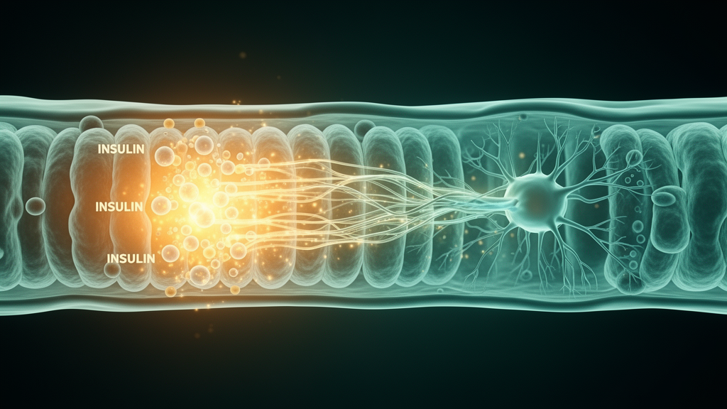
The study also traced the signaling routes behind these changes. After seven days of co-culture, the AKT and ERK pathways—two cascades downstream of insulin and growth factor receptors—were activated in both cell types. The ERK signaling pathway mediated TIMP1 upregulation in the endothelial cells. This connects the physical contact between pericytes and endothelial cells to insulin-sensitive intracellular signaling and, from there, to a concrete change in the barrier’s enzymatic environment.
Exosome analysis added another layer. BDNF was significantly enriched in exosomes isolated from the co-culture media on the abluminal side—the side facing the brain tissue—compared with exosomes from the solo cultures.
The model also captured a feature directly relevant to Alzheimer’s disease pathology. When pericytes were co-cultured with endothelial cells, the accumulation of toxic amyloid-beta 42 in the pericytes was reduced. This reduction occurred alongside lower expression of low density lipoprotein receptor-related protein 1 (LRP1), a receptor involved in clearing amyloid-beta from brain tissue. This observation ties the cell-cell interaction to reduced expression of LRP1, a receptor involved in clearing amyloid-beta from brain tissue, though the study did not demonstrate a full clearance mechanism in vivo.
The authors conclude that these findings provide mechanistic insights into blood-brain barrier disruption due to pericyte degeneration and highlight the role of insulin resistance at the barrier in causing cerebrovascular dysfunction in Alzheimer’s disease. Because the work was performed entirely in an in vitro co-culture system, the results describe mechanisms operating in human cells in a dish rather than confirmed processes in patients, and the stronger early-phase responses suggest timing matters in how these interactions unfold. Still, by linking pericyte-endothelial contact, insulin pathway activation, TIMP1-mediated MMP inhibition and amyloid-beta handling within a single human-cell model, the study offers a framework for understanding how the barrier’s supporting cells might protect—or fail to protect—the brain in Alzheimer’s disease.
Subject of Research: Endothelial-pericyte interactions, insulin signaling and blood-brain barrier dysfunction in Alzheimer's disease studied in a human in vitro co-culture model
Article Title: Endothelial-pericyte interactions activate insulin signaling and its implications for blood-brain barrier dysfunction in Alzheimer’s disease
Article References: Wang, Z., Veerareddy, V., Eberts, P. M., Azarin, S. M., & Kandimalla, K. K. (2026). Endothelial-pericyte interactions activate insulin signaling and its implications for blood-brain barrier dysfunction in Alzheimer’s disease. Fluids and Barriers of the CNS. https://doi.org/10.1186/s12987-026-00884-5
Image Credits: AI Generated
DOI: 10.1186/s12987-026-00884-5
Keywords: blood-brain barrier, pericytes, endothelial cells, insulin signaling, Alzheimer's disease, TIMP1, MMPs, amyloid-beta, exosomes, BDNF, LRP1, hiPSC-derived iBMECs
Cite Scienmag News
Cassandra Pierce. (October 28, 2026). Cell Talk at the Blood-Brain Barrier Switches On Insulin Signaling. Scienmag. https://scienmag.com/cell-talk-at-the-blood-brain-barrier-switches-on-insulin-signaling/
Cassandra Pierce. "Cell Talk at the Blood-Brain Barrier Switches On Insulin Signaling." Scienmag, 28 October 2026, https://scienmag.com/cell-talk-at-the-blood-brain-barrier-switches-on-insulin-signaling/. Accessed 29 October 2026.
Cassandra Pierce. "Cell Talk at the Blood-Brain Barrier Switches On Insulin Signaling." Scienmag. October 28, 2026. https://scienmag.com/cell-talk-at-the-blood-brain-barrier-switches-on-insulin-signaling/