


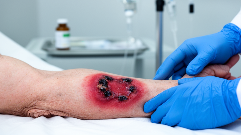
A cancer drug taken by thousands of patients with kidney cancer has been linked to a rare and dangerous skin condition that so closely mimics a flesh-eating bacterial infection that it nearly triggered unnecessary emergency surgery. In a case report published in the Journal of Cutaneous Immunology and Allergy, dermatologists at Hiroshima University Hospital in Japan describe how a 70-year-old man treated with the multi-kinase inhibitor cabozantinib developed severe skin necrosis that looked for all the world like necrotizing fasciitis, the rapidly progressive soft tissue infection colloquially known as flesh-eating disease. Only surgical exploration and a skin biopsy revealed the truth: the tissue death was driven not by an infection devouring his fascia, but by microscopic blood clots choking off blood supply to his skin, a complication apparently caused or worsened by the drug itself.
The case is believed to be the first reported instance of necrotizing cellulitis mimicking necrotizing fasciitis in a patient receiving a vascular endothelial growth factor inhibitor, a class of targeted cancer therapies that includes cabozantinib. The finding carries practical weight for clinicians worldwide, because distinguishing between the two conditions determines whether a patient must undergo radical, tissue-removing surgery or can be managed more conservatively with antibiotics and supportive care.
The patient’s story began 18 months before his crisis. He had been taking cabozantinib for advanced renal cell carcinoma, initially at 40 milligrams per day, a dose later cut in half because of hand-foot syndrome, a common side effect marked by painful redness and blistering of the palms and soles. When spinal metastases caused complete paralysis of his lower limbs, his oncologists raised the dose back to 40 milligrams per day. Three months after that increase, ulcers and swelling appeared on his lower legs. A month later, he developed fever and declining consciousness and was rushed to hospital.
What the team found on examination was alarming. His left lower leg was grossly swollen and covered in purpura, petechiae, erosions, and fluid-filled bullae. Laboratory results deepened the concern: his platelet count had collapsed to 9.5 × 10³ per microliter, his creatinine had climbed to 2.93 milligrams per deciliter, and inflammatory markers including C-reactive protein and procalcitonin were dramatically elevated. His Laboratory Risk Indicator for Necrotizing Fasciitis score came in at 9, well above the threshold that typically raises suspicion for the deadly infection. He was also in disseminated intravascular coagulation, a systemic clotting and bleeding derangement, and had underlying diabetes with chemotherapy-induced immunosuppression, both of which further heightened concern.
Yet the picture was not as straightforward as it seemed. Plain computed tomography showed inflammation confined largely to the subcutaneous tissue, with no gas formation and no deep fascial involvement, features that would be expected in true necrotizing fasciitis. When surgeons made an exploratory incision, they found full-thickness skin necrosis but no purulent discharge and no fat necrosis. The fascia beneath appeared viable and normally colored, and the finger test, in which a surgeon probes the plane between fascia and subcutis to see whether tissue peels away easily, was negative. Those findings argued against necrotizing fasciitis.
A skin biopsy delivered the decisive clue. Under the microscope, the dermis was studded with fibrin thrombi, microscopic clots plugging the small vessels that feed the skin, with no evidence of vasculitis. Blood and tissue cultures grew Streptococcus dysgalactiae, a streptococcal species capable of causing serious invasive infection. The team revised the diagnosis to necrotizing cellulitis with S. dysgalactiae sepsis and disseminated intravascular coagulation, and concluded that cabozantinib-associated microthrombi had caused the necrotizing fasciitis-like findings.
Cabozantinib is a multi-kinase inhibitor that targets the vascular endothelial growth factor receptor 2, the MET receptor tyrosine kinase, and the AXL receptor tyrosine kinase. By blocking these signaling pathways, it suppresses angiogenesis, the formation of new blood vessels that tumors rely on for growth and spread. But the same anti-angiogenic action injures the endothelial cells lining blood vessels throughout the body, and the drug is known to increase the risk of thromboembolism. The Hiroshima team speculates that this endothelial injury created a prothrombotic state that, when combined with a streptococcal infection and disseminated intravascular coagulation, produced microvascular thrombosis and tissue necrosis far out of proportion to what the infection alone could explain.
The distinction between necrotizing cellulitis and necrotizing fasciitis rests on the depth of tissue involvement. Necrotizing cellulitis affects the epidermis and subcutaneous tissue, while necrotizing fasciitis extends along the fascial planes, the tough connective tissue sheets that compartmentalize the body. The distinction is not academic: surgical debridement is mandatory for necrotizing fasciitis, which can kill within hours as infection races along fascial planes, but may not be required for necrotizing cellulitis, which can often be managed with antibiotics and conservative wound care. The LRINEC score, though elevated in this case, has limited specificity and can be raised in severe infections and inflammatory states such as sepsis and disseminated intravascular coagulation, and the clinical course here was not the rapid progression typical of necrotizing fasciitis.
Cabozantinib was discontinued, and the patient was treated with systemic antibiotics and anticoagulants. Despite extensive epidermal necrosis and ulceration, conservative management led to the formation of granulation tissue by day 42, sparing him from radical debridement. The authors emphasize that clinicians treating patients with vascular endothelial growth factor inhibitors should be aware that these therapies may cause severe cellulitis with thrombotic features that closely resemble necrotizing fasciitis, and that surgical and histopathological findings are essential to avoid unnecessary surgery and guide appropriate treatment.
Subject of Research: Cabozantinib-associated microthrombi causing necrotizing fasciitis-like findings
Article Title: Cabozantinib-associated microthrombi causing necrotizing fasciitis-like findings
Article References: Kawamoto, I., Matsuo, Y., Sakamoto, T., Kishi, M., Numata, T., Kan, T., & Tanaka, A. (2026). Cabozantinib-associated microthrombi causing necrotizing fasciitis-like findings. Journal of Cutaneous Immunology and Allergy, 9, Article 16659. https://doi.org/10.3389/jcia.2026.16659
Image Credits: AI Generated
Keywords: cabozantinib, microthrombi, necrotizing cellulitis, necrotizing fasciitis, VEGF inhibitor, renal cell carcinoma, Streptococcus dysgalactiae, Cabozantinib-associated, causing, necrotizing, fasciitis-like, findings
Cite Scienmag News
Nathaniel Bowman. (October 16, 2026). Cancer Drug Cabozantinib Linked to Rare Flesh-Eating Bacteria Mimic. Scienmag. https://scienmag.com/cancer-drug-cabozantinib-linked-to-rare-flesh-eating-bacteria-mimic/
Nathaniel Bowman. "Cancer Drug Cabozantinib Linked to Rare Flesh-Eating Bacteria Mimic." Scienmag, 16 October 2026, https://scienmag.com/cancer-drug-cabozantinib-linked-to-rare-flesh-eating-bacteria-mimic/. Accessed 16 October 2026.
Nathaniel Bowman. "Cancer Drug Cabozantinib Linked to Rare Flesh-Eating Bacteria Mimic." Scienmag. October 16, 2026. https://scienmag.com/cancer-drug-cabozantinib-linked-to-rare-flesh-eating-bacteria-mimic/


