


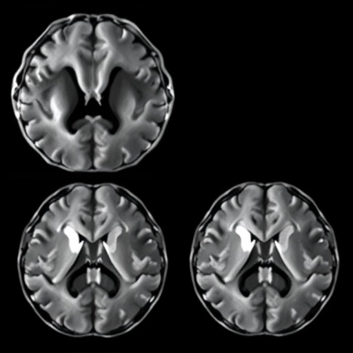
Parkinson’s disease may be entering a new era of prediction, in which the earliest clues to a patient’s future are found not inside a brain scanner, but in the clinical examination room. A study published in npj Parkinson’s Disease reports that baseline clinical features outperformed structural magnetic resonance imaging, or MRI, when researchers attempted to identify patients at risk of rapid cognitive and motor decline. The finding challenges a widely held assumption that increasingly detailed images of the brain will necessarily provide the most powerful forecast of how Parkinson’s disease will progress.
The research, led by Y. Wu, J.A. Santiago, T. Rundek and colleagues, focuses on one of the most urgent problems in neurology: Parkinson’s disease does not follow a single trajectory. Some people experience relatively gradual changes over many years, while others develop disabling movement problems, cognitive impairment or both at a much faster pace. At diagnosis, however, physicians often have limited tools for distinguishing these paths. This uncertainty affects treatment planning, clinical monitoring, caregiver preparation and the design of clinical trials, where researchers need to identify participants likely to show meaningful progression within a realistic timeframe.
MRI has long been viewed as a promising source of biological information. Structural MRI can measure the volume, thickness and shape of different brain regions, potentially revealing tissue loss associated with neurodegeneration. In principle, these anatomical signatures could provide an objective forecast of disease progression. Yet Parkinson’s disease is not simply a disorder of visible brain shrinkage. Its earliest and most consequential changes involve complex networks, neurotransmitter systems and microscopic cellular processes that may occur before large structural differences become detectable on routine scans.
The new study suggests that information gathered through standard clinical assessment may capture these processes more effectively than structural imaging alone. Baseline clinical features can include the pattern and severity of movement symptoms, age at assessment, cognitive performance, functional abilities and other measurable characteristics recorded when a patient first enters evaluation. Such observations are not merely descriptive. A patient’s balance, gait, speech, tremor, rigidity, response speed and performance on cognitive tests reflect the combined activity of multiple neural systems. They may therefore act as indirect but highly sensitive indicators of damage that a structural scan cannot yet resolve.
This distinction is technically important. Structural MRI primarily records anatomy: the distribution and volume of gray matter, white matter and other visible tissue compartments. It does not directly measure dopamine release, synaptic failure, inflammation, abnormal protein accumulation or the efficiency of communication between brain regions. Parkinson’s disease involves disruptions across these levels. A person may develop substantial functional impairment while their overall anatomical changes remain subtle, diffuse or inconsistent. Clinical testing, by contrast, samples the output of the entire nervous system, integrating many biological abnormalities into observable behavior.
The researchers’ conclusion does not mean that MRI is unhelpful or that brain imaging has no role in Parkinson’s disease. Instead, it indicates that structural MRI, when considered as a predictor in the context examined by the study, may not contain enough prognostic information to surpass a carefully collected clinical baseline. Imaging can still assist with diagnosis, help exclude other neurological conditions and contribute to research models when combined with more specialized techniques. Functional imaging, diffusion imaging, molecular scans and longitudinal measurements may reveal biological signals that a single structural scan misses. The central message is that the most sophisticated-looking measurement is not automatically the most informative one.
The finding could have immediate implications for medical care because clinical features are relatively accessible, inexpensive and repeatable. A neurological examination and standardized cognitive assessment can be performed in hospitals and clinics that do not have advanced imaging facilities. If validated in additional populations, clinical prediction tools could help physicians identify patients who need closer follow-up, earlier cognitive support or more intensive rehabilitation. They could also improve conversations with families by replacing vague expectations with a more individualized estimate of risk—although any prediction would still need to be presented as a probability rather than a certainty.
The result is especially relevant to the development of new treatments. Parkinson’s trials often face a major statistical challenge: participants progress at different speeds, making it difficult to determine whether an experimental therapy is genuinely altering the disease or whether the study population simply contains a mixture of rapid and slow progressors. A reliable baseline prediction model could allow investigators to balance treatment groups more precisely, enrich trials with participants likely to reach a defined clinical milestone and reduce the time required to detect meaningful differences. It could also help researchers test whether a therapy changes the expected course of decline rather than merely easing symptoms temporarily.
At the same time, the study highlights the limits of prediction in a biologically diverse disease. A model that performs well in one research cohort may be less accurate in another because of differences in age, disease duration, medication use, education, genetics, healthcare access or the way symptoms are measured. Clinical features can also change with treatment and may be influenced by conditions unrelated to Parkinson’s disease. For that reason, a prediction system must be tested across different hospitals, ethnic groups and stages of illness before it can be trusted for routine decisions. External validation, transparent reporting and regular recalibration will be essential.
The broader lesson is that medical progress does not always come from adding a more complicated instrument. In Parkinson’s disease, a structured record of how a person moves, thinks and functions at baseline may currently provide a clearer window into future decline than the anatomy visible on a conventional scan. The next generation of prognostic tools will likely combine both approaches, linking clinical observations with imaging, blood-based markers, genetics and digital measurements from smartphones or wearable sensors. By showing that baseline clinical features can outperform structural MRI, Wu and colleagues have redirected attention toward a practical but powerful idea: the patient’s living symptoms may be among the most information-rich signals available at the very beginning of the disease journey.
Cite Scienmag News
Diana Fleming. (August 19, 2026). Baseline clinical features outperform MRI in predicting rapid cognitive-motor decline in Parkinson’s. Scienmag. https://scienmag.com/baseline-clinical-features-outperform-mri-in-predicting-rapid-cognitive-motor-decline-in-parkinsons/
Diana Fleming. "Baseline clinical features outperform MRI in predicting rapid cognitive-motor decline in Parkinson’s." Scienmag, 19 August 2026, https://scienmag.com/baseline-clinical-features-outperform-mri-in-predicting-rapid-cognitive-motor-decline-in-parkinsons/. Accessed 3 September 2026.
Diana Fleming. "Baseline clinical features outperform MRI in predicting rapid cognitive-motor decline in Parkinson’s." Scienmag. August 19, 2026. https://scienmag.com/baseline-clinical-features-outperform-mri-in-predicting-rapid-cognitive-motor-decline-in-parkinsons/


