


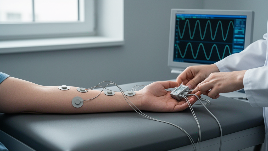
Surface electromyography has long promised clinicians a non-invasive window into the electrical activity of working muscles, a window that could one day help track recovery after stroke or other neurological and musculoskeletal injuries. Yet the technology has remained largely confined to laboratories, in part because its measurements are exquisitely sensitive to exactly where the electrodes sit on the skin. Move a sensor by even a centimetre and the recorded signal can change dramatically, making it nearly impossible to tell whether a difference between two recording sessions reflects genuine physiological change in a patient or simply sloppy repositioning of the hardware. A new study published in Annals of Biomedical Engineering tackles this problem head-on, presenting an algorithm that uses high-density electrode arrays to work out where the sensors have moved between sessions and to correct for that movement automatically.
The research, led by Fraser Douglas and colleagues at the University of British Columbia, rests on a simple but powerful idea: if you record muscle activity from a dense grid of 64 electrodes rather than a single pair, the spatial pattern of that activity acts like a fingerprint of the underlying muscle. Shift the grid and the fingerprint shifts with it. By comparing the spatial maps of muscle activity captured in one session with those from another, an optimisation algorithm can estimate the translation and rotation that occurred between the two recordings, without needing any external measurement of where the electrodes actually are.
To test the approach, the team recruited 11 healthy participants who performed a battery of exercises while wearing flexible 8-by-8 high-density sEMG arrays on four lower-limb muscles: the gastrocnemius medialis and tibialis anterior on the shank, and the semitendinosus and tensor fascia latae on the thigh. The exercise protocol included maximal isometric contractions specific to each muscle, 30 seconds of repeated sit-to-stand movements, and a ten-pace timed-up-and-go test, both dynamic tasks that feature routinely in stroke assessment sessions. After each round of exercises, the researchers took a 3D scan of the leg with the array still attached, then peeled the array off and reapplied it at a new location, shifted by up to 4.25 centimetres and rotated by up to 30 degrees in either direction. This process was repeated across four placement locations per muscle, generating hundreds of paired recordings in which the true change in electrode position was known from the scans.
The shift estimation algorithm itself works in three stages. First, the raw signals are filtered to remove noise and mains interference, then segmented into individual contractions. Next, three metrics are computed for every one of the 64 channels: the mean absolute pairwise correlation between each channel and its neighbours, which maps out regions of common underlying muscle activity; the mean frequency of the signal, which reflects how the frequency content of the EMG changes with electrode position; and the maximum envelope amplitude, a common time-domain measure related to force production. Finally, a gradient-descent-based global optimiser searches over all possible combinations of horizontal translation, vertical translation and rotation, seeking the shift that minimises the weighted error between the metric maps from the two sessions in their overlapping region. The weights were calibrated on a subset of 30 shift pairs, with the correlation maps proving the most informative contributor.
The results were striking. Across 263 isometric shift pairs, the algorithm improved the estimate of electrode location over the standard assumption that nothing had moved in 81.7 percent of cases. The median mean Euclidean distance between the estimated and true electrode positions fell from 3.00 centimetres under the no-shift assumption to 1.22 centimetres with the algorithm, and 37.6 percent of shifts were identified to within one centimetre, the spacing between adjacent electrodes in the array. Performance varied somewhat by muscle, with the tibialis anterior faring best, its shifts detected to within one centimetre in half of all cases. Notably, the researchers point out that certified prosthetist orthotists asked to locate a muscle belly by palpation in people with transtibial amputation managed to get within one centimetre of the ultrasound-identified location in only 28.8 percent of cases, suggesting the algorithm can match or exceed expert-guided placement precision.
Crucially, the team then showed that knowing where the electrodes moved actually matters for the measurements clinicians would want to make. They compared five standard sEMG features, including mean, median and peak frequency, integrated EMG and maximum envelope amplitude, across sessions using three strategies: comparing the closest electrodes as determined by the ground-truth 3D scans, comparing the closest electrodes as determined by the algorithm, and simply comparing the same physical electrodes before and after the shift, which is what current practice effectively does. For nearly all features, the same-electrode comparison produced significantly larger differences than either closest-electrode strategy. For the isometric maximum envelope amplitude, the mean relative difference rose from 15.9 percent when the algorithm’s correction was applied to 21.1 percent when it was not. The algorithm-corrected results were statistically indistinguishable from the ground-truth results, meaning the correction recovered essentially all of the accuracy that perfect knowledge of electrode position would provide.
The picture was more complicated for dynamic exercises. When the algorithm was applied to the sit-to-stand and timed-up-and-go data, it improved the shift estimate in only 63.1 and 56.3 percent of cases respectively, identifying the shift to within one centimetre in roughly 16 to 17 percent of cases. The researchers attribute this to the movement of muscles beneath the array during dynamic contractions, imprecise manual segmentation of contractions, and electrical crosstalk from neighbouring musculature. Even so, there was an encouraging exception: for the tibialis anterior during sit-to-stand, shifts were detected to within one centimetre in 32.3 percent of cases. That matters clinically because the tibialis anterior drives dorsiflexion, and its weakness after stroke causes drop foot, a hallmark impairment in which patients struggle to clear the foot during the swing phase of walking. Tracking that muscle’s recovery is among the most valuable things sEMG could offer stroke rehabilitation.
The authors are candid about the study’s limitations. All shifts were performed within a single data collection session lasting a few hours, rather than across the days or weeks that separate real clinical assessments, so performance over true multi-session intervals remains unconfirmed. The participants were healthy young adults, and the algorithm’s assumption that the limb can be approximated as a cylinder may not hold for amputees or people with reduced limb volume. The optimiser’s search space was bounded at four centimetres of translation and 30 degrees of rotation, though the team notes that 35 of 36 clinician placements in the earlier amputee study would have fallen within those bounds. There were also cases, about 16 percent, where the algorithm made the location estimate worse than doing nothing, often because it locked onto a spurious minimum at the edge of its search space. And because mean frequency and envelope amplitude serve both as inputs to the shift estimation and as clinical features, the researchers checked whether simply picking electrodes with the most similar feature values could substitute for resolving the shift; it could not, with only 6.3 percent of such electrodes for mean frequency falling within two centimetres of the true closest electrodes.
Looking ahead, the team plans to repeat the data collection with an older chronic stroke population, adding a second recording session two weeks after the first to test whether the approach generalises across genuine multi-session intervals and to a clinical population. They also hope to extend the framework to track muscle movement during dynamic contractions in real time, and to explore signal decomposition techniques that could keep the analysis stable in acute stroke, where motor unit recruitment patterns change rapidly. For now, the study demonstrates a concrete, quantified step toward making sEMG robust to the placement variability that has held it back, bringing the prospect of objective, sensor-based recovery tracking closer to the clinic and to remote assessment platforms where patients cannot rely on an expert to place their electrodes perfectly every time.
Subject of Research: An algorithm that quantifies and corrects electrode location changes in inter-session lower-limb surface electromyography recordings
Article Title: Improving the Clinical Utility of Lower-Limb Surface Electromyography (sEMG) by Quantifying and Correcting for Location Changes in Inter-Session Recordings
Article References: Douglas, F., Pei, M., Vu, Q. S., Le, L., & Kuo, C. (2026). Improving the Clinical Utility of Lower-Limb Surface Electromyography (sEMG) by Quantifying and Correcting for Location Changes in Inter-Session Recordings. Annals of Biomedical Engineering. https://doi.org/10.1007/s10439-026-04431-z
Image Credits: AI Generated
DOI: 10.1007/s10439-026-04431-z
Keywords: surface electromyography, high-density sEMG, electrode shift, stroke rehabilitation, muscle activity, biomedical engineering, signal processing, clinical assessment, wearable sensors, recovery tracking, lower-limb muscles, algorithm
Cite Scienmag News
Cassandra Pierce. (October 26, 2026). Algorithm Tracks Shifting Electrodes to Make Muscle Sensor Readings Reliable Across Sessions. Scienmag. https://scienmag.com/algorithm-tracks-shifting-electrodes-to-make-muscle-sensor-readings-reliable-across-sessions/
Cassandra Pierce. "Algorithm Tracks Shifting Electrodes to Make Muscle Sensor Readings Reliable Across Sessions." Scienmag, 26 October 2026, https://scienmag.com/algorithm-tracks-shifting-electrodes-to-make-muscle-sensor-readings-reliable-across-sessions/. Accessed 26 October 2026.
Cassandra Pierce. "Algorithm Tracks Shifting Electrodes to Make Muscle Sensor Readings Reliable Across Sessions." Scienmag. October 26, 2026. https://scienmag.com/algorithm-tracks-shifting-electrodes-to-make-muscle-sensor-readings-reliable-across-sessions/


