


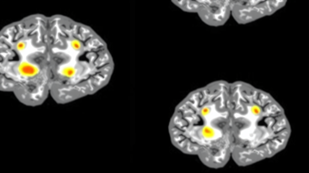
Glioblastoma has long been one of the most unforgiving diagnoses in medicine. It is the most common malignant brain tumor in adults and, by a wide margin, the most lethal, with a median survival of roughly 17 months after diagnosis. Surgeons can remove every piece of tumor visible to the eye and on the operating room imaging, patients can complete radiation and chemotherapy, and yet the cancer almost always comes back. That pattern of near-certain recurrence has shaped decades of research, and it is precisely the problem a new artificial intelligence system from the University of California, San Francisco and the University of Michigan is now taking aim at. In a study published September 25 in Science Advances and supported by the National Institutes of Health, the researchers describe an AI-based approach that can predict, with a high degree of accuracy, where a patient’s first recurrence is likely to appear, potentially opening a window to treat the tumor before it ever becomes visible on a standard MRI scan.
The clinical logic behind the work rests on a well-known but stubbornly difficult fact about glioblastoma biology: the disease rarely returns in a random location. Most patients experience recurrence at or close to the cavity left behind after the tumor is removed. That means the seeds of the next tumor are almost certainly already present in the tissue surrounding the resection site at the time of surgery, invisible to conventional imaging and often to the surgeon’s eye as well. If clinicians could identify which stretches of that surrounding tissue harbor infiltrating tumor cells, they could in principle act on that knowledge immediately, either by removing additional tissue during the initial operation or by directing focused therapies at the highest-risk zones before the disease re-establishes itself.
Turning that idea into a practical tool required solving a fundamental bottleneck in operating room pathology. The research team, led by first author Sanjeev Herr, MD, a postdoctoral research fellow at UC San Francisco, analyzed tissue samples collected during glioblastoma surgery from patients treated at UCSF Health, for whom the median time to recurrence was 5.5 months. Rather than relying on conventional pathology, which requires tissue to be fixed, processed with dyes and stains, and interpreted by a pathologist in a process that is both slow and labor-intensive, the team used a technique called stimulated Raman histology, or SRH. SRH produces microscopic images of fresh, unprocessed tissue in less than a minute, preserving the tissue in a near-native state and dramatically compressing the timeline from sampling to analysis.
Stimulated Raman histology works by exploiting the intrinsic vibrational signatures of the molecules inside tissue. When laser light interacts with lipids, proteins, and other cellular components, the scattered light carries chemical information that can be translated into images resembling traditional stained histology, all without a single drop of dye. That speed and chemical richness make SRH an ideal input for machine learning, because the raw optical data can be fed directly into an algorithm trained to recognize patterns that correlate with tumor infiltration. In this study, the researchers paired SRH imaging with FastGlioma, an AI system previously developed by investigators at UCSF and the University of Michigan, which scores tissue based on the degree of tumor infiltration it detects.
The scale of the study reflected the rigor needed to validate such a tool. The researchers developed their model using approximately 300 tissue samples drawn from 60 patients, then tested it separately on about 100 samples from a different set of 20 patients, keeping the training and testing data strictly apart. The central question was whether the AI’s assessment of infiltration, computed from fresh tissue in minutes, could predict where recurrent tumor would later emerge. The answer was encouraging on its own: the AI infiltration score alone performed about as well as conventional pathology at predicting which sampled areas would later develop recurrent disease. In other words, the algorithm extracted essentially the same prognostic signal from unprocessed tissue that the gold standard of surgical pathology extracts from stained slides, but in a fraction of the time.
The most striking results, however, came when the team layered additional information on top of the AI score. The researchers combined the infiltration measure with clinical, imaging, and molecular data and tested the resulting feature set across six different machine-learning models. The best-performing model was significantly more likely to distinguish between tissue sites that would and would not go on to develop recurrences than any single data type could manage alone. Notably, the AI measure of tumor infiltration proved to be the strongest individual predictor of recurrence in five of the six models, providing more predictive information than the tumor’s molecular characteristics, which are traditionally among the most powerful prognostic markers in glioblastoma. That finding suggests the spatial pattern of microscopic invasion, as read by the algorithm, captures something about recurrence risk that even genomic profiling misses.
Precision mattered as much as predictive power. A tool that says recurrence is coming somewhere in the general vicinity of the tumor cavity would be of limited use to a surgeon deciding where to extend a resection or where to aim a radiation boost. The researchers therefore tested how precisely the model could localize the future site of relapse, and it performed well at predicting whether cancer would return within 5 or 10 millimeters of the tissue that had been sampled. At those spatial scales, the difference between a predicted high-risk zone and an unremarkable one becomes actionable: five to ten millimeters is within the range a neurosurgeon can meaningfully consider when weighing how much additional tissue to remove, and it is squarely within the targeting capability of modern focal radiation techniques.
The therapeutic implications flow directly from that precision. Senior author Shawn Hervey-Jumper, MD, a neurosurgeon at UCSF Health and the Mitchel S. Berger, MD, endowed professor at the Weill Institute for Neurosciences, outlined two distinct pathways by which the predictions could change practice. For patients whose high-risk tissue lies in regions of the brain that can safely be resected, the AI guidance could support removing additional tissue during the initial surgery, clearing infiltrating cells before they have a chance to repopulate the cavity. For patients whose disease sits in parts of the brain that cannot be removed, the predicted sites of progression could instead be targeted with other treatments, such as higher-dose focal radiation or drugs infused directly into the tumor through a catheter placed through the skull. In both scenarios, the goal is the same: intervene while the residual disease is still microscopic and most vulnerable.
The system’s developers also see it functioning as a real-time decision support tool rather than a post-hoc analysis. Herr described the platform as having incredible potential to provide neurosurgeons with valuable real-time guidance during tumor removal, while also generating insights that guide subsequent treatment decisions. Because SRH imaging and AI scoring can be completed in under a minute on fresh tissue, the workflow could in principle fit inside a single operation, giving the surgical team infiltration maps while the patient is still on the table. That stands in sharp contrast to the current standard, in which definitive pathology results often arrive days after the operation, when the opportunity to act on them surgically has already passed.
The researchers deliberately focused their predictions on the first recurrence, a choice that reflects both scientific caution and clinical strategy. Patients with glioblastoma often receive experimental treatments later in the course of their disease, and those interventions can influence tumor growth in ways that make subsequent recurrences far more difficult to predict reliably. The first recurrence, arriving before most of those confounding therapies, offers the cleanest signal for validating a predictive model. It is also the recurrence that matters most strategically, as co-senior author Todd Hollon, MD, of the Machine Learning in Neurosurgery Laboratory at the University of Michigan, Ann Arbor, emphasized: the overall goal was to delay that first recurrence, and ultimately the team hopes that extending that window could translate into longer survival. For a disease in which median survival is measured in months and every extension of progression-free time carries real weight, a tool that converts fresh tissue, rapid optical imaging, and machine learning into a map of where the tumor will strike next represents one of the more concrete near-term applications of AI in neuro-oncology, and a potential shift from reacting to recurrence toward anticipating it.
Subject of Research: AI prediction of glioblastoma recurrence sites using stimulated Raman histology
Article Title: Treating a deadly brain tumor before it recurs? AI tool may help
Article References: Treating a deadly brain tumor before it recurs? AI tool may help. (n.d.). Original publication
Image Credits: AI Generated
DOI: Not provided
Keywords: glioblastoma, artificial intelligence, stimulated Raman histology, brain tumor recurrence, FastGlioma, neurosurgery, machine learning, tumor infiltration, focal radiation, UCSF, Science Advances, precision medicine
Cite Scienmag News
Nathaniel Bowman. (September 25, 2026). AI Model Predicts Where Glioblastoma Will Return Before MRI Can See It. Scienmag. https://scienmag.com/ai-model-predicts-where-glioblastoma-will-return-before-mri-can-see-it/
Nathaniel Bowman. "AI Model Predicts Where Glioblastoma Will Return Before MRI Can See It." Scienmag, 25 September 2026, https://scienmag.com/ai-model-predicts-where-glioblastoma-will-return-before-mri-can-see-it/. Accessed 6 October 2026.
Nathaniel Bowman. "AI Model Predicts Where Glioblastoma Will Return Before MRI Can See It." Scienmag. September 25, 2026. https://scienmag.com/ai-model-predicts-where-glioblastoma-will-return-before-mri-can-see-it/


