Scientists have documented, for the first time in full detail, how the sea star Asterias forbesi develops from a fertilized egg into a juvenile, and in doing so they have uncovered something unexpected: the same organ can be built through strikingly different sequences of steps, even among closely related sea stars. The study, published in Discover Developmental Biology, used brightfield microscopy and live imaging to chart every major stage of A. forbesi embryonic and larval development, an ecologically important North Atlantic species that, despite decades of research on its reproduction, behavior and role as a keystone predator, had never received a complete developmental description.
The research focused on a structure the authors call the hydro-vascular organ, or HVO. In all sea star larvae, a hollow tube of tissue called the coelom forms and eventually fills the larval body cavity. Because most of this organ goes on to become the adult water-vascular system, the network of fluid-filled canals and tube feet that sea stars use for locomotion, the team recently named it the hydro-vascular organ. Previous work in the model sea star Patiria miniata had shown that the HVO is essential: gene knockout experiments demonstrated that larvae lacking this organ do not survive. Its function in larvae is still debated, with hypotheses including buoyancy and excretion, but its importance is not in question.
The developmental account begins with the oocyte. A. forbesi oocytes are optically clear and spherical, arrested in prophase I with a large nucleus, the germinal vesicle, containing a prominent nucleolus. When the hormone 1-methyladenine was added to the culture medium, the oocytes resumed meiosis within twenty minutes, the nucleus broke down and the oocytes matured. After fertilization, the fertilization envelope rose from the oocyte surface, and the first cell divisions proceeded equally and synchronously, producing loosely bound blastomeres. As divisions continued, blastomeres became smaller and organized into a single monolayer epithelium surrounding a cavity, forming a pre-hatched blastula that rotated within the envelope before hatching to swim using beating cilia.
Gastrulation began at roughly 18 hours post fertilization, when the entire embryo elongated along its anterior-posterior axis and cells at the vegetal pole invaginated to form the archenteron, the future gut. The point of invagination marked the blastopore, the embryo’s first opening. The researchers observed that in every embryo examined, a single mesenchymal cell appeared before gastrulation started and moved into the blastocoel, an uncommon trait the authors suggest may be a polar body. As the gut reached the middle of the blastocoel, cells at its tip changed shape, acquired a mesenchymal phenotype and migrated out. Between about 40 and 44 hours, the tip of the gut expanded laterally to form two closed coelomic pouches, one on each side of the anterior gut, while the gut bent toward a ventral depression in the ectoderm and fused with it to form the mouth; the blastopore moved ventrally to become the anus.
Organogenesis followed in the early larva. The gut differentiated into a tripartite digestive system of esophagus, stomach and intestine, and a small rounded posterior enterocoel appeared on the posterior left side of the stomach, the site where primordial germ cells are thought to localize, though the authors note this will require germ cell markers to confirm. In about 10 percent of embryos, two posterior enterocoels were present. By day 3, the left coelom had elongated dorsally into a tube that fused with the ectoderm to form the hydropore, the only opening connecting the coelom to the outside environment. The larvae also displayed the characteristic ciliary bands of sea star bipinnaria larvae, composed of ciliated cells that form the locomotor apparatus, and two distinct cell populations were visible in the blastocoel: highly motile mesenchymal cells with long filopodia, and immobile granular cells embedded in the ectoderm. Both types were more abundant in the anterior half of the larva and more frequently associated with the ciliary band, a pattern quantified across 74 larvae from six batches.
As larvae matured into brachiolariae, the team documented features that set A. forbesi apart from the more commonly used P. miniata. A. forbesi larvae develop long arms, and fluorescent immunostaining revealed approximately 16 parallel muscle fibers running through the middle of each arm from the blastocoel to the tip, suggesting that the fast, forceful breaststroke-like swimming movements these larvae use are powered by muscle contraction. In P. miniata, by contrast, the short parallel muscle fibers were extensions of the dorsal muscles located underneath the arm ectoderm, not in the middle of the arms, a difference that reflects the fact that P. miniata larvae rely primarily on beating cilia rather than their arms for propulsion. Adult structures began appearing at week 3, when skeletal spicules formed on the stomach and the hydrocoel epithelium thickened and folded into five lobes, the rudiment of the primary podia. Distinctive settlement structures also developed, including a pair of brachiolar arms, a median ventral lobe, six pigmented adhesive papillae and an adhesive disk used to attach to the substratum during metamorphosis. Most larvae underwent metamorphosis in around three months, a process completed in about a week.
The central analytical contribution of the study is the division of HVO morphogenesis into four stages. Stage 1 began at the end of gastrulation, around 40 hours, when the tip of the gut bent outward on its left and right sides; by 44 hours, two coelomic pouches had budded off and begun elongating toward the posterior. In stage 2, the pouches detached from the foregut, and by 72 hours the right pouch stayed in the larval midline while the left moved dorsally and formed the hydropore canal. In stage 3, both pouches became tubes that elongated first posteriorly along the stomach, then anteriorly over the mouth. Quantification showed that the lumen area of the left tube was significantly larger than the right, a difference that persisted until the tubes merged, and the two tubes also differed in shape, with the right tube rounder during the first hours of organogenesis. In stage 4, the tubes reached the larval apex and merged anteriorly to form the single looped HVO, with a second connection forming posterior to the intestine around week 3. The organ proved highly muscular, with thick parallel muscle fibers around it, and live imaging showed the left tube twitching and contracting, suggesting the muscular layer helps pump fluid through the lumen.
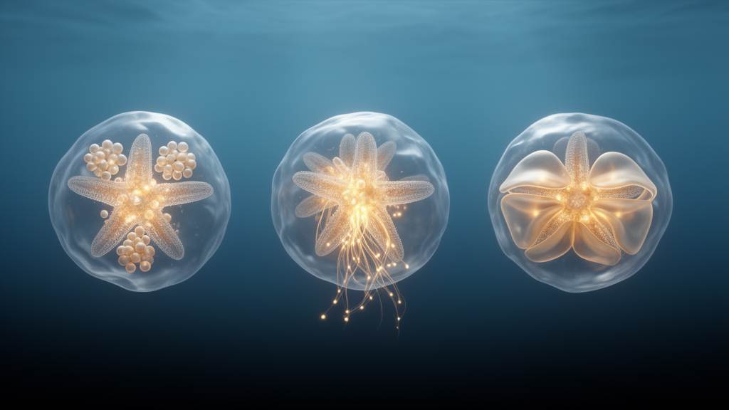
With A. forbesi characterized, the team compared HVO development across three species from three different sea star orders: A. forbesi (Forcipulatida) from the Northwest Atlantic, P. miniata (Valvatida) from the US Pacific coast, and Astropecten aranciacus (Paxillosida) from the Mediterranean. In all three, the HVO originates from the tip of the growing gut, but the elongation strategies diverge. In P. miniata, the coelomic pouches immediately acquire a tube shape and merge anteriorly above the mouth as early as day 3, before elongating posteriorly. In A. forbesi, the tubes first elongate posteriorly along the stomach and only then grow anteriorly to merge over the mouth. A. aranciacus follows a third strategy: the pouches form long, wide tubes that grow posteriorly, and the first connection between them is made posteriorly, around the intestine, before the connected tubes grow anteriorly. The authors note that their brightfield images of A. aranciacus, a species whose larval development had previously been described only through drawings, recapitulated that earlier source, and that further imaging is needed to define the specific epithelial changes observed at the archenteron tip in that species.
The comparison extends to other echinoderms. Sea urchin larvae also develop two coelomic pouches that bud from the anterior end of the foregut, but these tubes have constrictions along their length, never merge anteriorly, and form compartments. Holothurians develop pouches that never fuse into a single organ and do not make a rudiment. Sea stars, the authors conclude, are the only echinoderm larvae that build a full, looped organ that grows to fill the entire coelomic cavity. The finding that three sea star species from different orders arrive at similarly shaped final HVOs through three different developmental routes speaks to a broader question in developmental biology: how a conserved organ can form through distinct steps even within a single class of animals.
The study has clear limits that the authors themselves acknowledge. The work is descriptive, based on brightfield microscopy, live imaging and immunostaining of laboratory-cultured embryos at 18 degrees Celsius for A. forbesi and P. miniata and 15 degrees for A. aranciacus, and it does not identify the molecular mechanisms driving the different morphogenetic strategies. In P. miniata, previous work has shown that the FGF pathway drives tube elongation and the Wnt pathway regulates growth directionality, but whether these pathways play equivalent roles in A. forbesi remains an open question that genomic and transcriptomic investigations could address. The suggested role of the HVO in maintaining the shape of larval protrusions, and the hypothesis that arm movements are neurally controlled, both require further study. Still, the detailed staging and anatomical characterization now available for A. forbesi should prove useful for future research on reproduction, larval behavior and ecology, and the comparative framework establishes sea stars as a powerful system for asking how organs evolve their shapes.
Subject of Research: Comparative developmental staging of the sea star Asterias forbesi and the morphogenesis of its hydro-vascular organ
Article Title: Comparative analysis of Asterias forbesi development reveals distinct mechanisms of hydro-vascular organ formation across sea stars
Article References: Marc, T. K., Rodger, A., & Perillo, M. (2026). Comparative analysis of Asterias forbesi development reveals distinct mechanisms of hydro-vascular organ formation across sea stars. Discover Developmental Biology, 236(1), Article 4. https://doi.org/10.1007/s00427-026-00731-5
Image Credits: AI Generated
DOI: 10.1007/s00427-026-00731-5
Keywords: Asterias forbesi, sea stars, developmental biology, hydro-vascular organ, morphogenesis, larval development, coelom, Patiria miniata, Astropecten aranciacus, echinoderms, live imaging, organogenesis
Cite Scienmag News
Drew Townsend. (October 28, 2026). Sea star larvae build the same organ in three different ways, study finds. Scienmag. https://scienmag.com/sea-star-larvae-build-the-same-organ-in-three-different-ways-study-finds/
Drew Townsend. "Sea star larvae build the same organ in three different ways, study finds." Scienmag, 28 October 2026, https://scienmag.com/sea-star-larvae-build-the-same-organ-in-three-different-ways-study-finds/. Accessed 28 October 2026.
Drew Townsend. "Sea star larvae build the same organ in three different ways, study finds." Scienmag. October 28, 2026. https://scienmag.com/sea-star-larvae-build-the-same-organ-in-three-different-ways-study-finds/