


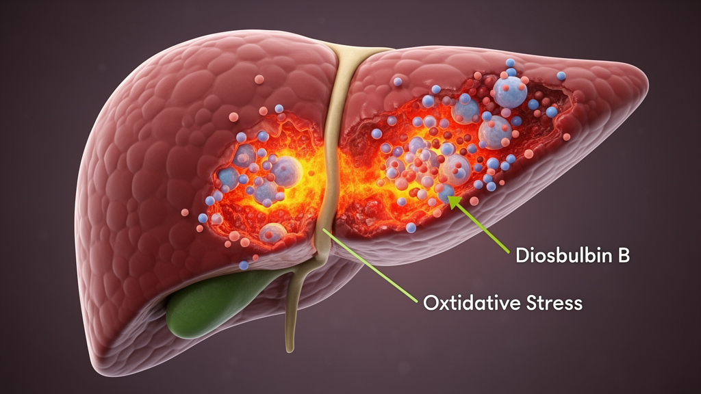
A furanoterpenoid compound found in a traditional Chinese medicinal plant appears to make a common chemotherapy drug harder on the liver, according to a new study in mice and in liver-derived cells. Diosbulbin B, a major hepatotoxic constituent of Dioscorea bulbifera L., was found to intensify liver injury markers in mice treated with doxorubicin, a widely used anthracycline chemotherapeutic whose clinical use is limited by cumulative organ toxicity. The work, published in Frontiers in Cell and Developmental Biology, combined serum biochemistry, histology, cell assays, bulk RNA sequencing and Western blotting to map how the two substances interact in hepatic tissue and in LO2 hepatocyte-like cells.
The research team, led by Yanqing Song of The First Hospital of Jilin University, set out to address a gap in herb–drug interaction research. While most natural-product studies around doxorubicin toxicity have focused on antioxidant or cytoprotective interventions that might shield organs from chemotherapy damage, far less attention has gone to toxic herbal constituents that could increase susceptibility to chemotherapy-associated injury. Earlier work had shown that Dioscorea bulbifera exposure delayed doxorubicin excretion and aggravated doxorubicin-induced cardiotoxicity and nephrotoxicity, and that a rhizome extract of the plant worsened pirarubicin-associated toxicity by affecting hepatic drug transporters. But those studies examined complex plant preparations and centered on the heart and kidney, leaving open whether purified diosbulbin B specifically aggravates doxorubicin-induced hepatotoxicity.
In the animal experiment, the researchers used specific pathogen-free male ICR mice aged 4 to 6 weeks, randomly assigned to four groups of eight animals each: a control group receiving saline and vehicle, a doxorubicin group receiving 3 mg/kg by intraperitoneal injection on days 1 and 3, a combined group receiving the same doxorubicin regimen plus 100 mg/kg diosbulbin B by oral gavage on the same days, and a diosbulbin B-only group. The animals were euthanized on day 9, and blood and liver tissue were collected for analysis. The doxorubicin dose was deliberately low and repeated, giving a cumulative 6 mg/kg, to induce measurable hepatotoxicity while limiting systemic mortality.
The results showed a clear pattern of enhanced injury with the combination. Compared with controls, both the doxorubicin and combined groups lost body weight, while the diosbulbin B-only group remained comparable to controls. Hematoxylin and eosin staining revealed inflammatory-cell infiltration and disruption of hepatic architecture in the doxorubicin group, with similar histopathological alterations in the combined group. Critically, serum alanine aminotransferase activity was higher in the combined group than in the doxorubicin-only group, and aspartate aminotransferase activity was highest of all in the combined group. The authors interpret these findings as suggesting that diosbulbin B co-treatment was associated with more severe doxorubicin-related liver injury under the experimental conditions, while cautioning that an animal model rather than clinical samples limits direct clinical extrapolation.
One of the more puzzling findings concerns redox chemistry. Rather than showing the classical signature of oxidative damage, the serum assays produced a non-classical pattern: glutathione levels rose in the doxorubicin and combined groups, with the highest level in the combined group; malondialdehyde levels fell in the treatment groups relative to controls; and superoxide dismutase activity decreased across all treatment groups. Serum lactate dehydrogenase showed no obvious difference among the four mouse groups. The authors argue that these changes indicate an altered redox-related biochemical status rather than direct evidence of a specific oxidative cell-death mechanism, and they warn against inferring oxidative stress from any single biochemical marker. They suggest the elevated glutathione may reflect an adaptive or compensatory response to toxic stress, noting that serum glutathione does not necessarily mirror intracellular or liver-tissue pools, and that the reduced malondialdehyde could relate to the sampling time point, altered lipid metabolism, or turnover of lipid peroxidation products.
In parallel, the team used LO2 human hepatocyte-like cells as a simplified in vitro model, treating them with doxorubicin at 1 or 2 micromolar, diosbulbin B at 100 or 200 micromolar, or combinations, for 24 or 48 hours. Doxorubicin reduced cell viability, and the combination groups showed lower viability than the corresponding doxorubicin-only groups, while diosbulbin B alone caused relatively limited changes at the tested concentrations. DAPI staining revealed condensed and irregularly shaped nuclei, most pronounced with the combination. JC-1 staining indicated more pronounced mitochondrial membrane depolarization with combined exposure than with doxorubicin alone, and lactate dehydrogenase release into the culture supernatant, a marker of membrane damage, was higher in the doxorubicin group than in controls and higher still in the combined group. Based on these results, the researchers selected 2 micromolar doxorubicin and 200 micromolar diosbulbin B for subsequent molecular assays.
Transcriptomic profiling added a genome-scale view. RNA sequencing of control, doxorubicin, and combined-treatment cells, with three biological replicates per group, showed distinct transcriptomic profiles by principal component analysis. In the doxorubicin-plus-diosbulbin B versus doxorubicin comparison, 1,221 differentially expressed genes were identified, with a striking asymmetry: only 130 genes were upregulated while 1,091 were downregulated. For context, the doxorubicin versus control comparison yielded 4,462 upregulated and 3,188 downregulated genes. Gene Ontology and Kyoto Encyclopedia of Genes and Genomes enrichment analyses pointed to terms and pathways related to transcriptional regulation, cell division, DNA repair, cellular response to DNA damage stimulus, double-strand break repair, homologous recombination, chromatin organization, mitotic spindle checkpoint signaling, cellular senescence, the cell cycle, and non-homologous end joining. Gene Set Enrichment Analysis highlighted mitotic spindle assembly checkpoint signaling, DNA double-strand break processing, and regulation of DNA damage response signal transduction. The authors emphasize, however, that enrichment analysis reflects coordinated gene-expression changes and should not be read as direct evidence of DNA damage or impaired repair.
Protein-level measurements sharpened the molecular picture. Western blotting showed that the phospho-PI3K/PI3K and phospho-AKT/AKT ratios were elevated in the doxorubicin and combined groups, with the highest levels in the combined group. P38 protein expression was higher in the combined group than in the other groups, although the study measured total p38 rather than phospho-p38, so the data describe increased expression rather than canonical p38 MAPK activation. TXNIP, IL-6, and IL-8 levels were increased in the doxorubicin and combined groups, again highest with the combination, and immunofluorescence staining for IL-6, p38, and NF-κB p65 showed the strongest signals in the combined group. Conversely, GADD45A and p53 protein levels were reduced in the combined group, consistent with the transcriptomic enrichment of cell-cycle and DNA damage-response pathways. The authors interpret the PI3K/AKT increases as possibly reflecting an adaptive or compensatory stress response, noting that without pathway inhibitors or genetic interventions the data cannot establish whether this signaling contributes causally to injury or is a secondary response.
The study’s limitations are stated plainly. No direct assays of lipid peroxidation-dependent cell death, ferroptosis, oxytosis, or necroptosis were performed; no direct DNA damage assays such as γ-H2AX staining, comet assays, or micronucleus assays were included; and no pathway-intervention experiments were conducted, leaving the roles of PI3K/AKT, p38, TXNIP, p53, and GADD45A associative. LO2 cells possess limited cytochrome P450 metabolic activity, so they cannot reproduce the metabolic activation-dependent hepatotoxicity of diosbulbin B, which is known to undergo cytochrome P450-mediated bioactivation into reactive intermediates that covalently bind cellular proteins. Pharmacokinetic interactions between the two compounds were not evaluated, and the stress-response proteins were validated only in cells, not in liver tissue. The mice were also young, at 4 to 6 weeks at treatment start. Within those bounds, the findings extend earlier concerns about Dioscorea bulbifera and anthracycline interactions to purified diosbulbin B and the liver, suggesting that this toxic herbal constituent may heighten susceptibility to doxorubicin-related hepatic injury and reshape hepatocellular stress-response programs, a caution of potential relevance for patients who combine herbal medicines with chemotherapy.
Subject of Research: Herb–drug interaction between diosbulbin B and doxorubicin in mouse and liver-cell models of hepatotoxicity
Article Title: Diosbulbin B aggravates doxorubicin-induced hepatotoxicity with PI3K/AKT signaling and redox-inflammatory stress responses
Article References: Xie, X., Xing, Y., Liu, X., Zhai, J., & Song, Y. (2026). Diosbulbin B aggravates doxorubicin-induced hepatotoxicity with PI3K/AKT signaling and redox-inflammatory stress responses. Frontiers in Cell and Developmental Biology, 14, 1970882. https://doi.org/10.3389/fcell.2026.1970882
Image Credits: AI Generated
DOI: 10.3389/fcell.2026.1970882
Keywords: diosbulbin B, doxorubicin, hepatotoxicity, herb–drug interaction, Dioscorea bulbifera, PI3K/AKT signaling, oxidative stress, mitochondrial membrane potential, LO2 cells, transcriptomics, p53, inflammation
Cite Scienmag News
Nathaniel Bowman. (October 27, 2026). Herbal compound diosbulbin B worsens doxorubicin liver injury in mice. Scienmag. https://scienmag.com/herbal-compound-diosbulbin-b-worsens-doxorubicin-liver-injury-in-mice/
Nathaniel Bowman. "Herbal compound diosbulbin B worsens doxorubicin liver injury in mice." Scienmag, 27 October 2026, https://scienmag.com/herbal-compound-diosbulbin-b-worsens-doxorubicin-liver-injury-in-mice/. Accessed 27 October 2026.
Nathaniel Bowman. "Herbal compound diosbulbin B worsens doxorubicin liver injury in mice." Scienmag. October 27, 2026. https://scienmag.com/herbal-compound-diosbulbin-b-worsens-doxorubicin-liver-injury-in-mice/


