


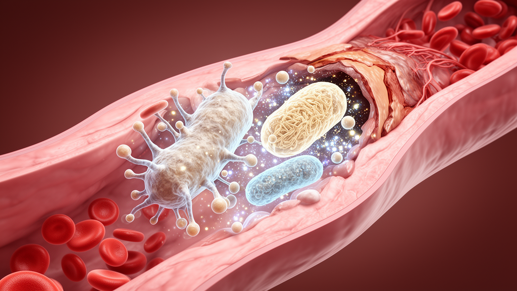
A 2026 review concludes that disturbed mitochondrial quality control in plaque macrophages is an active part of atherosclerosis, linking damaged-organelle clearance, altered mitochondrial dynamics, oxidative stress, inflammation, and foam-cell formation into a self-reinforcing disease process. The evidence comes mainly from macrophages and mouse models, with the strongest experimental results showing that manipulating mitophagy or mitochondrial fission can alter plaque size and stability. Most proposed treatments remain preclinical, and the review warns against treating macrophage mitochondrial dysfunction as a simple, uniform pathway.
The proposed mechanism begins with mitochondria failing to maintain the balance among mitophagy, fusion and fission, mitochondrial biogenesis, and mitochondrial DNA homeostasis. When damaged mitochondria are not removed, their electron transport chains become less efficient, reactive oxygen species accumulate, and mitochondrial DNA can escape into the cytosol. These events can activate the cGAS-STING pathway, the NLRP3 inflammasome, and other innate immune sensors, increasing IL-1β-associated sterile inflammation and contributing to foam-cell formation. The review frames mitochondrial quality-control imbalance as both a consequence of the plaque environment and a possible driver of lesion development, a causal question that current evidence cannot yet fully resolve.
Macrophages are well placed to amplify this process because they are the predominant immune cells in atherosclerotic plaques. Uptake of oxidized low-density lipoprotein imposes substantial metabolic stress, while plaque hypoxia and inflammation disturb mitochondrial function. Under less pathological conditions, macrophages rely heavily on mitochondrial oxidative phosphorylation and fatty-acid oxidation to preserve lipid homeostasis and anti-inflammatory activity. Persistent oxidative stress, impaired fusion, excessive fission, and defective mitophagy can shift their metabolism toward glycolysis. Accumulating succinate and citrate, increased mitochondrial reactive oxygen species, and stabilization of HIF-1α can then promote NLRP3 activation and a more inflammatory macrophage phenotype.
Impaired mitophagy provides one of the clearest experimental links between mitochondrial quality control and plaque disease. Loss of PINK1 or Parkin prevents efficient recognition and clearance of dysfunctional mitochondria, allowing electron leakage, mitochondrial DNA release, and inflammasome activation to increase. In ApoE-deficient mice, genetic loss of these mitophagy mediators accelerates lesion development and produces less stable plaques, whereas restoring macrophage mitophagy limits lesion growth. These findings support a functional role rather than a purely statistical association, but most experiments have used global genetic manipulation or systemic drugs. Macrophage-conditional studies with detailed measurements of necrotic cores, collagen, and defined macrophage populations remain limited.
Mitochondrial dynamics provide a second intervention point. Excessive fragmentation can compromise respiratory-chain activity, elevate mitochondrial reactive oxygen species, and increase susceptibility to inflammatory signaling. In high-fat-diet-fed ApoE-deficient mice, the Drp1 inhibitor Mdivi-1 reduced plaque area, NLRP3 activation, and M1-associated polarization; it also suppressed oxidized-LDL-induced inflammation in THP-1-derived macrophages through the mitochondrial reactive oxygen species–NLRP3 axis. The interpretation is nevertheless context-dependent. Drp1-dependent fission can also protect macrophages from mitochondrial DNA and ZBP1-driven inflammation after myocardial infarction, showing that inhibiting a fission pathway cannot be assumed to have the same effect in every vascular setting.
Mitochondrial DNA abnormalities offer a further possible upstream influence. Atherosclerosis-associated mitochondrial variants may be homoplasmic, present in every copy of the genome, or heteroplasmic, present in only a proportion of copies. Hybrid cell experiments showed that mitochondria carrying a single pro-atherosclerotic mutation produced defective mitophagy and an “intolerant” inflammatory response, in which repeated cytokine challenges failed to decline. Editing out the m.15059G>A mutation restored normal mitophagy and cytokine tolerance. Separately, elevated METTL4 was associated with mitochondrial DNA modification that suppressed MT-ATP6 expression, impaired respiratory-chain function, and promoted mitochondrial DNA release. The METTL4 inhibitor pemetrexed delayed atherosclerosis in an animal model, but the delivery challenge for mitochondrial editing remains substantial.
The inflammatory consequences can form a feedback loop between mitochondrial dysfunction and macrophage polarization. Mitochondrially damaged, highly inflammatory macrophages can produce TNF-α and IL-1β, which further disrupt mitochondrial function and sustain tissue injury. Mitochondrial repair or enhanced mitophagy may favor a less inflammatory, M2-like state in the simplified framework used by the review. That sequence, however, should not be treated as universal. Single-cell analyses of mouse and human plaques identify inflammatory monocyte-derived macrophages, TREM2-high foamy macrophages, and resident-like populations, and often find that non-foamy cells constitute the more inflammatory group. Mitochondrial quality-control measurements for these specific plaque subsets are still scarce.
Several intervention classes have improved outcomes in preclinical systems but differ sharply in their evidence and translational limitations. Rapamycin, metformin, and urolithin A have been used to enhance mitophagy through mTOR- or AMPK-SIRT1-related signaling, although their optimal dosing and long-term effects on human autophagy remain unresolved. Mdivi-1 and the mitochondrial protective peptide SS-31 target fission and membrane stability, but Mdivi-1 has context-dependent effects and potential off-target activity. MitoQ and Mito-TEMPO scavenge mitochondrial reactive oxygen species; MitoQ has improved endothelial function in a small randomized human trial, but broader cardiovascular antioxidant trials have not consistently shown benefit. Colchicine, canakinumab, and MCC950 act downstream through inflammation rather than correcting the mitochondrial defect itself.
Macrophage-selective delivery may be necessary if mitochondrial therapies are to move from experimental models into patients. Nanoparticle systems based on lipids or high-density-lipoprotein-like structures are being investigated to carry proteins, peptides, drugs, small interfering RNAs, and microRNAs to plaque macrophages. In theory, this could concentrate treatment in pathogenic subsets while limiting systemic toxicity from antioxidants, fission inhibitors, or metabolic modulators. Inhibiting glycolysis could also restore oxidative phosphorylation and an anti-inflammatory phenotype, but current approaches lack macrophage selectivity and raise concerns about systemic toxicity. Mitochondrial hydrogen sulfide delivery, lipid-based nanocarriers, and mitochondrial genome editors similarly remain at the in-vitro or preclinical stage.
The review’s most important qualification is that the evidence does not yet establish a universal therapeutic sequence from mitochondrial damage to inflammation, foam cells, and plaque rupture. Much of the literature is cross-sectional, so it remains unclear whether mitochondrial abnormalities precede atherosclerosis or arise from established lesions. RAW264.7 cells and ApoE-deficient mice also reproduce only parts of human plaque biology. Resolving the field will require longitudinal interventions, subset-resolved mitochondrial profiling in human plaques, and direct comparisons of mitochondrial manipulations in macrophages, vascular smooth muscle cells, and endothelial cells within the same animal model. The current evidence nevertheless identifies mitochondrial quality control as a plausible inflammatory and metabolic hub in atherosclerosis, with mitophagy restoration, dynamic-balance correction, antioxidant protection, and targeted delivery offering testable routes for future investigation rather than established clinical treatments.
Subject of Research: Macrophage mitochondrial quality control in atherosclerosis
Article Title: Mechanisms and targeted intervention strategies of macrophage mitochondrial quality control imbalance in the progression of atherosclerosis
Article References: Zhili, G., & Xiaoyu, Z. (2026). Mechanisms and targeted intervention strategies of macrophage mitochondrial quality control imbalance in the progression of atherosclerosis. Frontiers in Physiology, 17, 1975785. https://doi.org/10.3389/fphys.2026.1975785
Image Credits: AI Generated
DOI: 10.3389/fphys.2026.1975785
Keywords: atherosclerosis, macrophages, mitochondrial quality control, mitophagy, mitochondrial dynamics, mitochondrial DNA, NLRP3 inflammasome, foam cells, plaque stability, preclinical therapeutics, Mechanisms, targeted
Cite Scienmag News
Juliet Wilcox. (October 22, 2026). Review links macrophage mitochondrial imbalance to atherosclerosis progression. Scienmag. https://scienmag.com/review-links-macrophage-mitochondrial-imbalance-to-atherosclerosis-progression/
Juliet Wilcox. "Review links macrophage mitochondrial imbalance to atherosclerosis progression." Scienmag, 22 October 2026, https://scienmag.com/review-links-macrophage-mitochondrial-imbalance-to-atherosclerosis-progression/. Accessed 22 October 2026.
Juliet Wilcox. "Review links macrophage mitochondrial imbalance to atherosclerosis progression." Scienmag. October 22, 2026. https://scienmag.com/review-links-macrophage-mitochondrial-imbalance-to-atherosclerosis-progression/


