


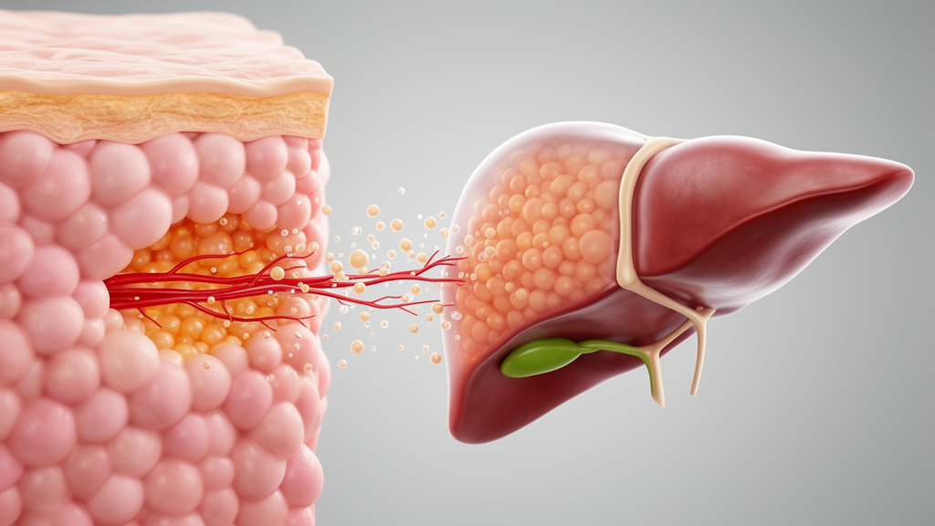
A 2026 narrative review argues that metabolic dysfunction-associated steatohepatitis, or MASH, in many people with type 2 diabetes is more faithfully understood as the hepatic expression of systemic adipose tissue dysfunction than as a disease that begins independently in the liver. The authors, Salvatore Corrao and Christiano Argano, organize the proposed mechanism into a continuous sequence: fat tissue loses its capacity to safely store energy, becomes inflamed and hormonally disordered, and sends excess lipids and inflammatory signals toward the liver. The resulting combination of insulin resistance, impaired fuel switching and hepatic lipotoxicity can turn fatty accumulation into inflammatory liver injury. The review presents this as a biologically coherent but hypothesis-generating framework, not a settled causal conclusion or a diagnostic rule.
Adipose tissue is portrayed as an active metabolic and immune organ rather than a passive reservoir. In its healthy state, subcutaneous fat can accommodate surplus energy by expanding adipocytes and recruiting new cells, thereby shielding the liver and other organs from circulating fatty acids. That buffering capacity can deteriorate when the tissue grows under chronic positive energy balance. Enlarged adipocytes may outstrip their blood supply, producing local hypoxia, oxidative stress, cell death and extracellular-matrix remodeling. These changes reduce the tissue’s ability to take up and safely retain lipid while increasing the release of fatty acids and inflammatory mediators. The result is a functional failure that need not track total body fat: one person may have substantial obesity but relatively preserved metabolic health, whereas another may develop severe insulin resistance and liver disease despite a leaner body.
The location of dysfunctional fat matters because different depots do not behave alike. Visceral adipose tissue drains directly into the portal vein, delivering concentrated fatty acids and adipokines to hepatocytes before those substances undergo systemic dilution. Intermuscular, epicardial, perivascular, perirenal and pancreatic fat also differ in cellular origin, secretory profile and metabolic effect. The review therefore argues that MASH reflects not simply how much fat a person has, but where that fat is located and how effectively it functions. It also cites lipodystrophy as an extreme demonstration of this principle: people with very little functional adipose tissue can develop severe insulin resistance and marked hepatic steatosis because they cannot safely store lipid. A genetically influenced, individual limit for safe adipose expansion may help explain why people with similar body sizes develop different metabolic outcomes.
Dysfunctional adipose tissue changes both its immune environment and its endocrine signaling. Obesity-associated macrophage accumulation can organize around dying adipocytes, while cytokines such as tumor necrosis factor-α, interleukin-6 and monocyte chemoattractant protein-1 sustain inflammation and interfere with insulin signaling. The authors distinguish “adipoflammation,” describing chronic inflammation within the fat depot, from “metaflammation,” referring to a low-grade inflammatory spillover into the circulation. This immune-metabolic feedback can become self-reinforcing: inflammation reduces safe lipid storage, greater ectopic lipid flux intensifies insulin resistance, and worsening insulin resistance further destabilizes adipose tissue. Endocrine signals change at the same time. Adiponectin, which promotes fatty-acid oxidation, restrains lipogenesis and has anti-inflammatory and liver-protective actions, generally falls as adipose dysfunction advances. Leptin rises with fat mass but can lose central metabolic effectiveness in insulin resistance, while resistin and other mediators are associated with insulin resistance and inflammation.
These changes produce a selective form of insulin resistance that helps explain how glucose and lipid disturbances can coexist. In the insulin-resistant liver, insulin may inadequately suppress gluconeogenesis while its lipogenic signaling remains active, allowing compensatory hyperinsulinemia to increase hepatic fat synthesis rather than restrain it. Lipid intermediates inside hepatocytes, including diacylglycerols and ceramides, can impair insulin-receptor signaling through pathways that include protein kinase C-ε. At the whole-body level, the authors connect this selective resistance with metabolic inflexibility, the impaired ability of tissues to switch between glucose and fatty-acid oxidation according to whether an organism is fed or fasting. That flexibility is difficult to measure directly, and the review notes that some apparent switching defects under experimental clamps reflect glucose-disposal rates rather than an independent biomarker. Even so, the model describes a coherent fuel imbalance: adipose tissue releases lipid it can no longer store, insulin resistance redirects substrate toward synthesis and ectopic deposition, and impaired switching prevents tissues from disposing of the surplus efficiently.
Because the liver receives nutrients from the gut and visceral fat products through the portal circulation, it becomes the most conspicuous site where this systemic disturbance becomes visible. Human isotope studies cited in the review attribute the largest share of stored hepatic triglyceride to circulating fatty acids released by adipose lipolysis, with de novo lipogenesis contributing a disproportionately large share in metabolic disease and dietary fat contributing less. Stored triglyceride is not necessarily the most damaging component, however. When incoming lipid exceeds the hepatocyte’s storage and oxidation capacity, reactive intermediates such as diacylglycerols, ceramides and incomplete oxidation products disrupt membranes, organelles and insulin signaling. Mitochondrial and endoplasmic-reticulum stress can then generate oxidative stress and inflammatory signals. Steatosis thus appears in this framework as a saturated hepatic buffer, while steatohepatitis reflects lipotoxic and immunometabolic injury once that buffer has been overwhelmed.
The progression from fat accumulation to inflammation is not presented as a simple, universally linear sequence. Lipotoxic intermediates, oxidative stress, organelle dysfunction, immune activation and altered gut-derived signals can act together on a susceptible liver. Microbial products and metabolites enter through the portal circulation, while intestinal barrier changes may increase the translocation of substances such as lipopolysaccharide. These can activate pattern-recognition receptors, including Toll-like receptor 4, on hepatic macrophages and add to the injury produced by lipid overload. Persistent hepatocyte damage then evokes a wound-healing response involving hepatic stellate cells and extracellular-matrix deposition, which contributes to fibrosis. The causal direction also runs back toward the liver: steatotic and inflamed hepatic tissue can reduce insulin clearance, sustain gluconeogenesis and release hepatokines such as fetuin-A and selenoprotein P, worsening glucose regulation and insulin resistance elsewhere.
Responses to interventions that improve systemic metabolic health provide indirect support for the framework. The review notes histological improvements in steatohepatitis with weight loss achieved through dietary energy restriction and physical activity, as well as with thiazolidinediones that improve insulin sensitivity and adiponectin signaling, incretin-based therapies that reduce adiposity and improve metabolic control, and metabolic surgery that produces sustained reductions in dysfunctional fat mass. These observations are consistent with an important upstream contribution from adipose dysfunction, but treatment response cannot prove a single disease origin: some drugs also act directly on the liver, and improving weight, insulin sensitivity or inflammation may influence several pathways at once. Likewise, the review’s proposed “dysfunctional fat immuno-inflammatory disease” is explicitly an interpretative construct with no diagnostic criteria, staging system or coding purpose; its value is organizational, bringing endocrine, immune and lipid-handling abnormalities under one research framework.
The model is principally intended for the common phenotype of metabolic dysfunction-associated steatotic liver disease associated with obesity, visceral adiposity and type 2 diabetes. It is less comprehensive for lean MASH, in which genetic variants including PNPLA3 and TM6SF2 may carry proportionally greater weight, and for patients with advanced fibrosis despite modest metabolic disturbance, whose disease may reflect greater intrinsic hepatic or fibrogenic susceptibility. Gut signaling, circadian disruption, genetic predisposition, bile-acid pathways, mitochondrial dysfunction and inherent hepatocyte susceptibility can all modify the process, and the relative contributions of adipose dysfunction and these alternatives cannot yet be quantified precisely. The review itself is a narrative synthesis of literature identified through PubMed and Google Scholar from January 2000 to June 2026, supplemented by reference-list searches; it is not a systematic review or a prospective test of the adipose-centered hypothesis. Its central proposal is consequently testable rather than proven: future studies could ask whether measures of adipose function predict MASH, fibrosis progression or treatment response independently of liver fat, body mass index and conventional risk factors. If confirmed, the fatty liver seen in type 2 diabetes would be not merely a hepatic diagnosis, but a visible sign of a disorder that begins in the adipose organ and then feeds back through the liver.
Subject of Research: Adipose tissue dysfunction as an upstream driver of MASH in type 2 diabetes
Article Title: Adipose tissue dysfunction and the liver: reframing MASH as the hepatic expression of systemic metabolic dysfunction
Article References: Corrao, S., & Argano, C. (2026). Adipose tissue dysfunction and the liver: reframing MASH as the hepatic expression of systemic metabolic dysfunction. Frontiers in Endocrinology, 17, 1920879. https://doi.org/10.3389/fendo.2026.1920879
Image Credits: AI Generated
DOI: 10.3389/fendo.2026.1920879
Keywords: MASH, type 2 diabetes, adipose tissue dysfunction, insulin resistance, metabolic inflexibility, hepatic steatosis, lipotoxicity, adipokines, adipose inflammation, gut-liver axis, Adipose, tissue
Cite Scienmag News
Daisy Hatcher. (October 22, 2026). Type 2 diabetes fatty liver may begin in dysfunctional adipose tissue. Scienmag. https://scienmag.com/type-2-diabetes-fatty-liver-may-begin-in-dysfunctional-adipose-tissue/
Daisy Hatcher. "Type 2 diabetes fatty liver may begin in dysfunctional adipose tissue." Scienmag, 22 October 2026, https://scienmag.com/type-2-diabetes-fatty-liver-may-begin-in-dysfunctional-adipose-tissue/. Accessed 22 October 2026.
Daisy Hatcher. "Type 2 diabetes fatty liver may begin in dysfunctional adipose tissue." Scienmag. October 22, 2026. https://scienmag.com/type-2-diabetes-fatty-liver-may-begin-in-dysfunctional-adipose-tissue/


