


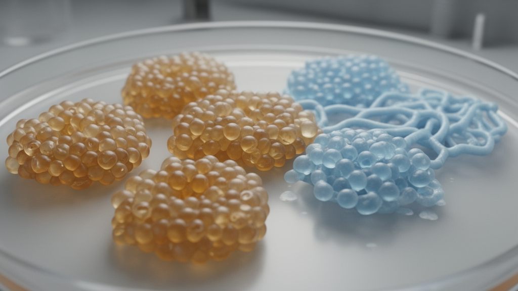
Adult pancreatic acinar cells can be kept in a recognizable, functioning state outside the body, then steered through an injury-associated transformation and partially redirected toward their original identity, according to a mouse study. The experiments establish a staged culture system in which different signals appear to initiate and sustain acinar-to-ductal metaplasia, the expansion of metaplastic cells, and their subsequent redifferentiation. The work also shows that the oncogenic KPC background facilitates conversion and destabilizes redifferentiation, while leaving it uncertain whether the results apply to human cells or to tumors in a living animal.
Acinar cells normally produce digestive enzymes and are held in a differentiated state by a transcriptional program involving factors such as Ptf1a, Rbpjl, Mist1/Bhlha15 and Nr5a2. Injury or oncogenic signaling can override that program, suppressing secretory genes and giving cells ductal or progenitor-like features. This process, called acinar-to-ductal metaplasia, or ADM, can resolve after acute injury but can persist under oncogenic Kras signaling and progress toward pancreatic ductal adenocarcinoma. A central unresolved question was whether cells that have undergone sustained metaplasia can reacquire an acinar identity rather than merely being replaced by residual acinar cells that survived.
To address that question, the researchers first screened culture conditions intended to preserve freshly isolated adult mouse acinar cells, which normally lose their identity within days. A defined maintenance medium containing nicotinamide, inhibitors of c-Jun N-terminal kinase and platelet-derived growth factor receptor, and the Notch inhibitor DAPT preserved compact acinar clusters for at least seven days. The cells retained expression of Amylase and Cpa1, showed little Krt19, and continued to release amylase in response to the secretagogue ceruletide. Although secretory-enzyme expression fell soon after plating, it subsequently stabilized, suggesting that the culture captured a functional acinar state rather than merely a static expression profile.
Adding epidermal growth factor to this stable baseline was sufficient to trigger metaplastic conversion. Over seven days, EGF reduced Amylase and Cpa1 while increasing proliferative and ductal-associated markers, and gefitinib, an EGFR inhibitor, opposed those changes. Cultures treated with EGF continued to show ductal and proliferative features over 20 days, whereas untreated cells maintained in the defined medium remained stable. These results identify EGF/EGFR signaling as an initiation cue for metaplastic conversion in this system, although the experiment did not establish that EGFR is the only route into ADM or that it operates identically in every tissue context.
The researchers then developed an expansion medium, ECCA, combining EGF with the Wnt agonist CHIR-99021 and the compounds CP-673451 and A83-01. Individual acinar cells labeled by the Mist1 lineage reporter developed into spheroids and then large cystic organoids. After seven days, 89.1% of the organoids carried the acinar lineage label, while organoids formed even when cells were diluted to densities at which individual colonies were widely separated, supporting an origin from single cells. The expanding organoids expressed ductal and progenitor-associated markers and retained little Amylase. Component-withdrawal experiments suggested that EGF acted mainly during the initial phase of organoid formation, whereas CHIR-99021 supported a broader growth period.
Single-cell RNA sequencing showed that this expansion followed an ordered trajectory. Lineage-restricted cells moved from an acinar state through an intermediate population that coexpressed Cpa1 and the ductal marker Krt19, then into predominantly ductal-like states by days five to seven. Pseudotime analysis placed these states along a single path, with a transient Reg3b-high intermediate and a progressive loss of acinar genes alongside later accumulation of Muc1 and Krt19. When projected onto published single-cell datasets from pancreatitis and Kras G12D models, the cultured cells mapped to corresponding acinar, ADM and metaplastic regions. Transcription-factor regulon activity followed the same temporal cascade in all three datasets, indicating that the culture reproduced conserved features of in vivo metaplasia without its stromal, immune, vascular or neural environment.
Expanded organoids retained evidence of acinar redifferentiation potential. After transfer to a staged differentiation medium, they compacted from cystic structures into grape-like clusters, re-expressed Amylase and Cpa1, reduced Krt19 and proliferation markers, and recovered measurable amylase secretion and ceruletide responsiveness. However, the maintenance medium alone was insufficient. The differentiation protocol required an initial four-day phase containing transient glucocorticoid receptor activation with dexamethasone and IGF1 receptor inhibition with picropodophyllin, followed by a maintenance phase in which those two components were withdrawn. Continuous exposure lowered ductal and proliferative markers but produced weaker late induction of acinar genes than the staged treatment, consistent with an initiation cue rather than a sustained requirement.
Pharmacological tests supported roles for both pathways. Dexamethasone induced canonical glucocorticoid receptor targets, its effect was reduced by the receptor antagonist mifepristone and reproduced by betamethasone, and the IGF1R inhibitor linsitinib reproduced the effect of picropodophyllin while added IGF1 opposed it. Yet the response depended on cellular context: dexamethasone reduced Ptf1a in freshly isolated acinar cells, and dexamethasone combined with IGF1R inhibition reduced Cpa1, unlike the expanded organoids. Redifferentiation was also incomplete and weakened after serial passage. The investigators could not exclude selection or persistence of residual acinar-like cells, and the study did not directly assess cell polarity, ultrastructure, survival or apoptosis.
The KPC mouse genotype, carrying Kras G12D, mutant Trp53 and Pdx1-Cre, altered both ends of the pathway. Wild-type acini required exogenous EGF for efficient organoid formation, and gefitinib almost completely blocked expansion. KPC acini formed large cystic organoids even without EGF, and EGFR inhibition reduced but did not abolish the response, indicating that the oncogenic background partially bypassed dependence on exogenous EGF. Because KPC combines two altered genes and the pancreas is remodeled before culture, the experiment cannot separate the contributions of mutant Kras and mutant p53 or establish that either acts autonomously in adult acinar cells. The pan-Kras inhibitor BI-2865 and the Kras G12D-selective inhibitor MRTX1133 reduced organoid outgrowth in KPC cultures, with MRTX1133 showing greater selectivity for that genotype.
Post-expansion redifferentiation was also less stable in KPC organoids. Amylase and Cpa1 rose only transiently before declining, and this response became weaker over successive passages, while KPC cultures retained more cystic organoids than wild-type cultures. MRTX1133 increased endpoint acinar-marker expression and strengthened Amylase staining while reducing Krt19, but it did not restore the sustained response seen in wild-type organoids. Taken together, the mouse ex vivo experiments show that acinar plasticity can be separated into controllable phases: EGFR-associated signaling initiates conversion, Wnt-associated support sustains expansion, and a transient combination of glucocorticoid receptor activation and IGF1R inhibition initiates redifferentiation. They also distinguish the capacity to reacquire acinar features from durable restoration of a fully mature acinar cell, providing a platform for testing the signals that govern each stage without claiming that the same mechanism resolves ADM in patients.
Subject of Research: Stage-specific regulation of acinar-cell plasticity in mouse pancreatic organoids
Article Title: Acinar‐Derived Organoids Reveal Stage‐Specific Control of Acinar Plasticity
Article References: Hou, Y., Li, S., Li, C., Lin, Z., He, D., Yang, L., Fu, L., Zhang, Q., Zhou, T., Yao, Y., & Yin, X. (2026). Acinar‐Derived Organoids Reveal Stage‐Specific Control of Acinar Plasticity. Advanced Science, Article e78350. https://doi.org/10.1002/advs.78350
Image Credits: AI Generated
DOI: 10.1002/advs.78350
Keywords: pancreatic acinar cells, organoids, acinar-to-ductal metaplasia, cell plasticity, EGFR signaling, redifferentiation, KRAS, single-cell RNA sequencing, Acinar, Derived, Reveal, Stage
Cite Scienmag News
Nathaniel Bowman. (October 20, 2026). Mouse Acinar Organoids Reveal Stage-Specific Control of Cell Plasticity. Scienmag. https://scienmag.com/mouse-acinar-organoids-reveal-stage-specific-control-of-cell-plasticity/
Nathaniel Bowman. "Mouse Acinar Organoids Reveal Stage-Specific Control of Cell Plasticity." Scienmag, 20 October 2026, https://scienmag.com/mouse-acinar-organoids-reveal-stage-specific-control-of-cell-plasticity/. Accessed 20 October 2026.
Nathaniel Bowman. "Mouse Acinar Organoids Reveal Stage-Specific Control of Cell Plasticity." Scienmag. October 20, 2026. https://scienmag.com/mouse-acinar-organoids-reveal-stage-specific-control-of-cell-plasticity/


