


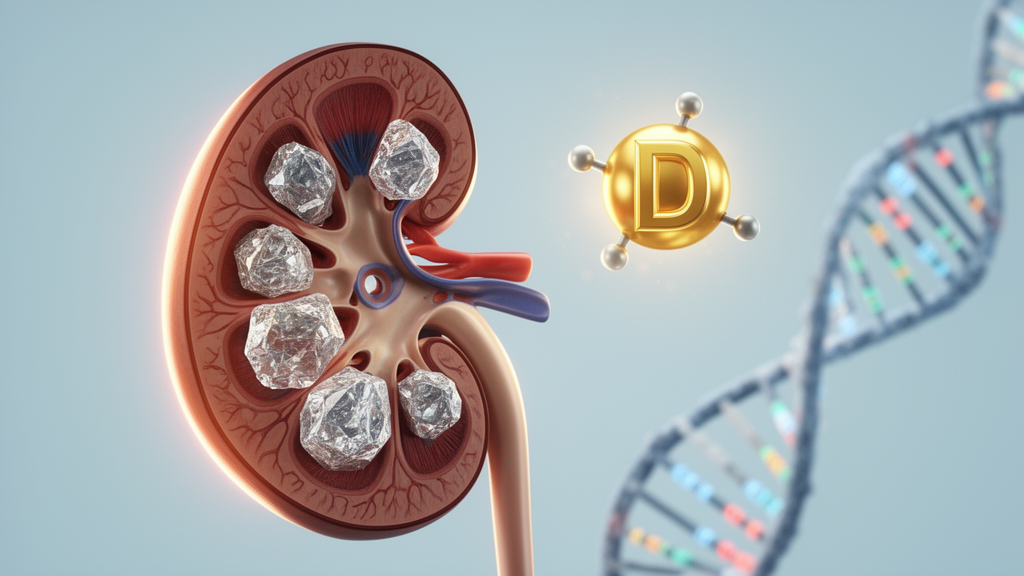
Kidney stones are one of the most painful and increasingly common conditions in modern medicine, and a new study from India suggests that the risk of developing them may hinge on an unexpected combination of factors: the level of vitamin D circulating in the blood and the specific genetic architecture of the vitamin D receptor. The research, published in Molecular Biology Reports, examined 267 patients with kidney stone disease and 296 carefully screened healthy controls from a genetically homogeneous population in northeastern India, and its findings challenge the simplistic view that vitamin D is either purely protective or purely harmful for stone formation. Instead, the study reveals that the interplay between serum vitamin D status and inherited variation in the vitamin D receptor gene can dramatically modify disease susceptibility, with some genetic variants amplifying risk while others appear protective.
The research team, led by Sanjenbam Yaiphaba Meitei of Manipur University, chose their study population with deliberate scientific precision. The Meitei people of Manipur represent a genetically homogeneous group, meaning that the confounding effects of population stratification, which often plague genetic association studies, are minimized. This homogeneity allowed the investigators to detect subtle genetic signals that might be obscured in more admixed populations. All participants underwent metabolic screening to rule out underlying systemic conditions that could independently influence stone formation, ensuring that the case-control comparison reflected genuine differences in vitamin D biology rather than secondary metabolic disturbances.
The methodological approach combined biochemical measurement with molecular genetics. Serum 25-hydroxyvitamin D, the standard clinical marker of vitamin D status, was quantified in all participants. Genomic DNA was extracted from blood samples, and three well-characterized polymorphisms in the vitamin D receptor gene were genotyped using polymerase chain reaction followed by restriction fragment length polymorphism analysis. These polymorphisms, known as ApaI, TaqI, and FokI, are restriction enzyme recognition sites that mark functional variation in the receptor gene. The FokI polymorphism is particularly interesting because it alters the translation initiation codon of the receptor protein, potentially producing a receptor variant with different transcriptional activity. The ApaI and TaqI variants, located in the 3′ untranslated region of the gene, may influence messenger RNA stability and receptor expression levels.
When the researchers analyzed each polymorphism individually, the results were initially disappointing. None of the three VDR variants showed a statistically significant association with kidney stone disease on its own. This finding aligns with the mixed results of previous meta-analyses, which have reported inconsistent associations between individual VDR polymorphisms and urolithiasis across different populations. However, the study’s most striking result emerged from the biochemical data: elevated serum vitamin D was strongly associated with disease risk, with an odds ratio of 12.55 and a 95 percent confidence interval spanning 4.70 to 33.54, a p-value below 0.001. This means that individuals with higher circulating vitamin D levels faced more than twelve times the odds of having kidney stones compared to those with lower levels.
To translate this biochemical association into a clinically useful threshold, the researchers performed receiver operating characteristic analysis, a statistical technique that evaluates the diagnostic accuracy of a continuous biomarker. The analysis identified an optimal vitamin D cut-off of 35.84 nanograms per milliliter, with an area under the curve of 0.816. An AUC of this magnitude indicates good discriminatory power, meaning that serum vitamin D levels above this threshold reliably distinguish stone patients from healthy controls in this population. The cut-off is notable because it falls within the range often considered sufficient or even optimal for bone health, raising the provocative possibility that vitamin D levels adequate for skeletal integrity may nonetheless promote urinary stone formation in genetically susceptible individuals.
The haplotype analysis represented the study’s most sophisticated genetic approach. Rather than examining each polymorphism in isolation, the researchers constructed haplotypes, which are combinations of alleles at multiple loci that are inherited together on the same chromosome. This approach captures the combined functional effect of linked genetic variants. The analysis identified the GCC haplotype as a significant risk factor for kidney stone disease, with an odds ratio of 4.08 and a 95 percent confidence interval of 2.47 to 6.72. In contrast, the GCT haplotype was strongly protective, with an odds ratio of 0.21 and a confidence interval of 0.10 to 0.46. These findings demonstrate that the combined genetic signature of the VDR gene, rather than any single variant, substantially modifies disease susceptibility. The GCC haplotype carries more than four times the risk, while GCT reduces risk by nearly 80 percent.
Linkage disequilibrium analysis, which measures the non-random association of alleles at different loci, provided additional context for the haplotype findings. The three polymorphisms showed patterns of linkage disequilibrium consistent with their physical proximity on chromosome 12, where the VDR gene resides. This non-random association means that certain allele combinations occur together more frequently than would be expected by chance, creating the haplotype blocks that the researchers identified as risk or protective signatures. The biological interpretation is that these haplotypes may represent functional units of the VDR gene, where the combined effect of multiple variants on receptor expression or activity differs from the sum of individual variant effects.
The gene-environment interaction analysis was the study’s crowning achievement. Using the additive interaction model developed by Rothman and refined by Andersson and colleagues, the researchers evaluated whether the combined effect of serum vitamin D status and VDR genotype exceeded the sum of their individual effects. The results were remarkable. Positive interactions were detected between elevated serum vitamin D and the ApaI GT genotype, as well as between elevated vitamin D and the FokI CC and CT genotypes. This means that individuals carrying these genetic variants face disproportionately higher risk when their vitamin D levels are elevated, compared to what would be predicted from the additive effects of each factor alone. In contrast, the TaqI variants showed antagonistic interactions, meaning that the presence of certain TaqI alleles actually buffered the risk associated with high vitamin D levels.
The biological plausibility of these interactions rests on the fundamental role of the vitamin D receptor in calcium homeostasis. When vitamin D binds to its receptor, the activated complex translocates to the nucleus and regulates the expression of genes involved in calcium absorption in the intestine and calcium reabsorption in the kidney. Elevated circulating vitamin D, combined with a receptor variant that has enhanced transcriptional activity, could drive excessive calcium transport, increasing urinary calcium concentration and promoting calcium oxalate crystallization. Conversely, a receptor variant with reduced activity might attenuate this effect even when vitamin D levels are high. The antagonistic interaction observed with TaqI variants supports this model, suggesting that certain receptor configurations protect against the lithogenic potential of elevated vitamin D.
The clinical implications of this study extend beyond the immediate findings. The research suggests that integrating biochemical and genetic markers may improve risk assessment for kidney stone disease and support precision prevention strategies tailored to individual genetic profiles. Rather than applying universal recommendations for vitamin D supplementation or restriction, clinicians may eventually be able to identify patients whose genetic constitution makes them particularly vulnerable to the lithogenic effects of high vitamin D levels. The study also highlights the value of studying genetically homogeneous populations, where the signal-to-noise ratio in genetic association analyses is enhanced. As kidney stone disease continues to rise globally, driven by dietary changes, climate warming, and increasing obesity, understanding the molecular mechanisms that govern individual susceptibility becomes ever more urgent. This study provides a template for future investigations that combine biochemical measurement, haplotype analysis, and gene-environment interaction modeling to unravel the complex etiology of this common and painful condition.
Subject of Research: Genetics
Article Title: Serum vitamin D and VDR genetic variation in kidney stone disease: haplotype and gene–environment interaction analyses in a genetically homogeneous population
Article References: Meitei, S. Y., Yumnam, M., Singh, A. R., Pebam, E., Konjengbam, H., Shantajit, N., & Paonam, S. S. (2026). Serum vitamin D and VDR genetic variation in kidney stone disease: haplotype and gene–environment interaction analyses in a genetically homogeneous population. Molecular Biology Reports, 53(1), Article 1734. https://doi.org/10.1007/s11033-026-12949-7
Image Credits: AI Generated
DOI: 10.1007/s11033-026-12949-7
Keywords: vitamin D, kidney stones, VDR gene, haplotype, gene-environment interaction, urolithiasis, genetics, precision medicine, Serum, vitamin, genetic, variation
Cite Scienmag News
Juliet Wilcox. (October 18, 2026). Vitamin D Levels and Receptor Gene Variants Combine to Drive Kidney Stone Risk. Scienmag. https://scienmag.com/vitamin-d-levels-and-receptor-gene-variants-combine-to-drive-kidney-stone-risk/
Juliet Wilcox. "Vitamin D Levels and Receptor Gene Variants Combine to Drive Kidney Stone Risk." Scienmag, 18 October 2026, https://scienmag.com/vitamin-d-levels-and-receptor-gene-variants-combine-to-drive-kidney-stone-risk/. Accessed 18 October 2026.
Juliet Wilcox. "Vitamin D Levels and Receptor Gene Variants Combine to Drive Kidney Stone Risk." Scienmag. October 18, 2026. https://scienmag.com/vitamin-d-levels-and-receptor-gene-variants-combine-to-drive-kidney-stone-risk/


