


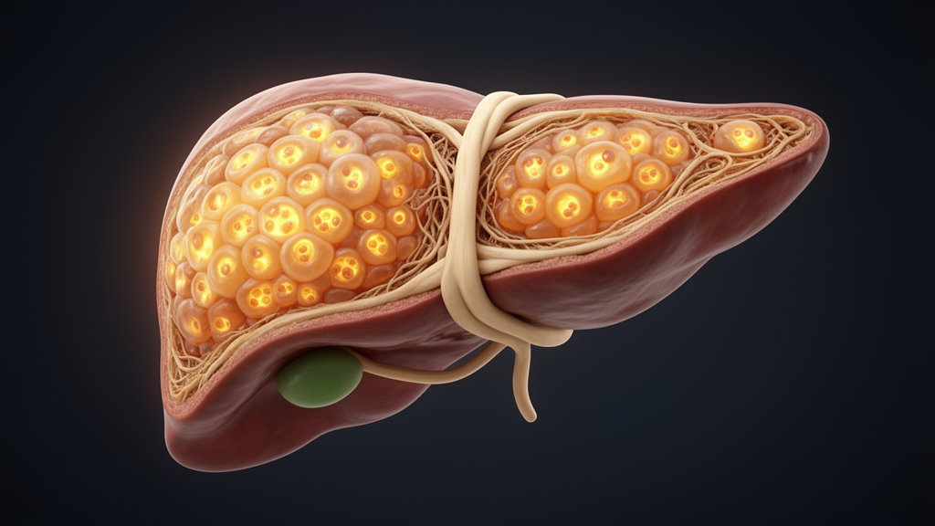
When the liver is scarred by cirrhosis, its working tissue is steadily replaced by useless connective tissue, and the organ must fight to rebuild itself. A new study published in Comparative Cytogenetics examines one of the least understood weapons in that fight: cellular, or somatic, polyploidy, the condition in which individual cells carry multiple complete sets of chromosomes. Working with both rats and humans, the researchers quantified exactly how much of the liver’s regenerative growth can be attributed to polyploidy, compared with the two better-known mechanisms of cell proliferation and cell enlargement. Their results reveal a striking difference between the two species, and they suggest that the polyploid state of liver cells is far more than an evolutionary curiosity.
Polyploidy comes in two fundamentally different forms. Generative polyploidy arises when a genomic mutation during meiosis produces gametes with extra chromosome sets, and it is inherited by every cell of the resulting organism. This whole-genome duplication has played a particularly important role in the evolution of plants and animals, although in animals it is much rarer than in plants and appears mainly in invertebrates and in vertebrates near the base of the evolutionary tree, chiefly fish and amphibians. In mammals, generative polyploidy is almost entirely absent, a fact thought to be connected with the genetic mechanism of sex determination. The single known exception is the red vizcacha rat from Argentina, the only recorded tetraploid mammal.
Mammals, however, display the second form: somatic polyploidy, which develops only in particular tissues or individual cells and is not inherited. In organs such as the liver and the heart, polyploid cells can make up a substantial share of the cell population. Typically the ploidy of these cells does not exceed the octoploid level, meaning eight sets of chromosomes, but in pathological conditions hepatocytes and heart muscle cells can reach 32-fold, 64-fold or higher ploidy. The number and ploidy levels of such cells vary strongly between individuals and are formed anew at certain stages of postnatal life through incomplete cell divisions.
The mechanics of this process are elegant. Alternating rounds of acytokinetic mitoses, in which a cell’s nucleus divides but the cell itself does not, produce binucleate cells, while so-called bimitoses double the chromosome content again: a diploid cell with amount of DNA denoted 2c becomes binucleate, then progresses through 4c and 8c states and beyond. Multipolar mitoses also contribute to the formation of polyploid hepatocytes. The details differ between mammalian species, but every pathway begins from the same starting pool of mononucleate diploid hepatocytes. At the cellular level, the most visible consequence of polyploidy is an increase in cell size, which in plants often produces gigantism; in animals, polyploid organisms usually remain the same body size as their diploid relatives and simply have fewer cells.
Why would liver cells adopt this strategy at all? The prevailing hypothesis is that hepatocyte polyploidisation evolved as a genetic mechanism of adaptation to the damaging effects of xenobiotics, the foreign chemicals that arrive with food. Because each extra chromosome set carries a full copy of every gene, polyploid cells change their metabolic indicators in accordance with gene dosage, both normally and under pathological conditions. Yet while polyploidy at the organismic level is recognised as a powerful engine of speciation, allowing new species to colonise habitats inaccessible to diploid ancestors, its role at the tissue level has remained largely obscure. That gap is what the new study set out to close, by measuring the contribution of polyploidy to normal and reparative liver growth in the rat, Rattus norvegicus, and in humans, Homo sapiens, and comparing it with proliferation and hypertrophy.
The experimental context is cirrhosis of various causes, a widespread human and animal disease in which functioning liver parenchyma is replaced by connective tissue. During the development of cirrhosis, the number of hepatocytes, the cells that perform the liver’s many functions, falls by 28 percent in rats and by twofold in humans, differences the authors report as highly significant. These profound disruptions to the organ’s architecture and metabolism trigger a powerful regenerative response: hepatocyte proliferation and hypertrophy both increase by roughly 25 percent compared with the normal state, in rats and humans alike. The question was which cellular processes actually account for the liver mass gained during this repair.
To find out, the team used a combined cytochemical method that determines the DNA content and dry mass of individual hepatocytes, allowing each cell to be assigned to a ploidy class. In normal rats, the modal hepatocyte is tetraploid: about 82 percent of cells fall into the 4c class, with smaller fractions of binucleate diploid cells and higher-ploidy cells, and an average ploidy of 4.46c. In rats with carbon tetrachloride-induced cirrhosis, the distribution shifts markedly. The proportion of binucleate diploid hepatocytes declines, while cells of high ploidy become more common, including a new appearance of 8c×2 cells, and the average ploidy rises by 14.8 percent. Humans present a different baseline: the normal human liver is dominated by mononucleate diploid 2c cells, which make up nearly 90 percent of the population, with an average ploidy of just 2.21c. In cirrhotic human livers, that average still climbs, by 15.8 percent, as diploid cells give way to binucleate and higher-ploidy forms.
The quantitative breakdown of growth mechanisms is where the two species truly diverge. Using formulae that partition changes in liver mass among proliferation, polyploidisation and hypertrophy, the researchers calculated that in rats, hypertrophy accounts for about 18 percent of the mass increase during cirrhosis development, but the dominant contributors are processes involving DNA synthesis and increased cell number: normal mitotic proliferation accounts for 66 percent and polyploidisation for 16 percent. In humans, the picture is entirely different. Reparative growth of the cirrhotic human liver was found to be due solely to the mitotic divisions of small diploid hepatocytes, with the calculated contributions of polyploidisation and hypertrophy coming out slightly negative, at minus 7.3 and minus 3.9 percent respectively. In other words, the human liver repairs itself in cirrhosis almost entirely by multiplying its remaining diploid cells, while the rat liver also leans heavily on making its existing cells bigger and more polyploid.
That intense proliferation of diploid hepatocytes in the human cirrhotic liver carries a warning. The authors note that it may indicate the transformation of the liver parenchyma into hepatocellular carcinoma, a possibility highlighted by several recent studies they cite. A regenerative program that depends on rapid division of the small diploid cell pool may come at the cost of pushing that pool toward malignant transformation, whereas the rat strategy of adding chromosome sets within existing cells avoids mass cell division. This contrast gives the species difference practical as well as evolutionary significance.
The study’s overall conclusion is twofold. Somatic polyploidy does make a significant contribution to the normal postnatal growth and the reparative growth of the liver, in rats and in humans, confirming that this tissue-level phenomenon is functionally meaningful rather than incidental. At the same time, the most significant contribution to increasing liver mass, both during ordinary development and during regeneration, comes from normal mitotic divisions of mononucleate diploid hepatocytes. Polyploidy, on this evidence, is a substantial supporting player rather than the lead: it buffers the damaged liver, enlarges and reinforces existing cells, and varies with pathology, but the workhorse of liver regeneration remains the humble diploid hepatocyte dividing in the classical way. For researchers hoping to coax cirrhotic livers into safer repair, understanding when and why hepatocytes choose extra chromosomes over extra cells may prove an important piece of the puzzle.
Subject of Research: The contribution of somatic cellular polyploidy to normal and regenerative liver growth in cirrhotic rats and humans
Article Title: The role of cellular polyploidy in the regeneration of the cirrhotic liver in rats and humans
Article References: Bezborodkina, N. N., Brodsky, V. Y., & Kudryavtsev, B. N. (2024). The role of cellular polyploidy in the regeneration of the cirrhotic liver in rats and humans. Comparative Cytogenetics, 18, 51-57. https://doi.org/10.3897/compcytogen.18.121459
Image Credits: AI Generated
DOI: 10.3897/compcytogen.18.121459
Keywords: polyploidy, liver regeneration, cirrhosis, hepatocytes, somatic polyploidisation, cell proliferation, hypertrophy, Rattus norvegicus, Homo sapiens, ploidy, comparative cytogenetics, hepatocellular carcinoma
Cite Scienmag News
Drew Townsend. (October 18, 2026). How Polyploid Liver Cells Help the Cirrhotic Rat Liver Regenerate. Scienmag. https://scienmag.com/how-polyploid-liver-cells-help-the-cirrhotic-rat-liver-regenerate/
Drew Townsend. "How Polyploid Liver Cells Help the Cirrhotic Rat Liver Regenerate." Scienmag, 18 October 2026, https://scienmag.com/how-polyploid-liver-cells-help-the-cirrhotic-rat-liver-regenerate/. Accessed 18 October 2026.
Drew Townsend. "How Polyploid Liver Cells Help the Cirrhotic Rat Liver Regenerate." Scienmag. October 18, 2026. https://scienmag.com/how-polyploid-liver-cells-help-the-cirrhotic-rat-liver-regenerate/


