


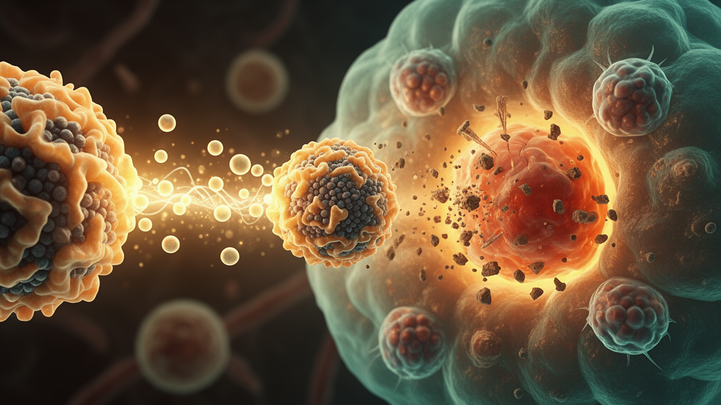
Glioblastoma remains one of the most stubborn enemies in modern oncology. Even with maximal surgical resection followed by radiotherapy and chemotherapy, most patients face a grim prognosis, and the outlook has barely shifted in decades. Two formidable barriers stand in the way: the blood-brain barrier, which blocks most therapeutic agents from reaching the tumor, and a deeply immunosuppressive tumor microenvironment that renders glioblastoma a classic immunologically “cold” tumor, largely unresponsive to immunotherapy. Now, a research team reporting in Materials Today Bio has engineered a biomimetic nanoplatform that tackles both problems at once, using a cloak of microglial cell membrane to ferry a cargo of carbon monoxide donor, manganese ions, and the pyroptosis effector Gasdermin E directly into brain tumors, where it triggers a cascade of mitochondrial destruction, inflammatory cell death, and immune activation.
The nanosystem, dubbed M@GM, is built around a manganese carbonyl (Mn2(CO)10) core loaded with recombinant GSDME protein and subsequently wrapped in membranes harvested from BV2 microglial cells, the resident immune cells of the central nervous system. The choice of microglial membrane is strategic: these membranes carry native protein compositions and immune-evasive properties that help the nanoparticles slip past the blood-brain barrier and home in on glioma tissue. Transmission electron microscopy revealed uniform spherical particles with a clear core-shell architecture after coating, while elemental mapping showed manganese signal colocalized with the nitrogen, sulfur, and phosphorus signals derived from the membrane, confirming successful encapsulation. Coomassie Brilliant Blue staining demonstrated that the protein band profile of the coated particles closely matched that of the native microglial membrane, indicating that the biological camouflage was preserved during fabrication.
Physicochemical characterization showed that M@GM nanoparticles have a hydrodynamic diameter predominantly in the range of 40 to 80 nanometers, averaging approximately 60 nanometers, with a zeta potential comparable to that of the native microglial membrane. X-ray photoelectron spectroscopy confirmed the presence of characteristic Mn 2p, O 1s, C 1s, and Cl 2p peaks. The loading contents of GSDME and MnCO were quantified by UV-vis spectrophotometric mass-balance methods at 7.2 percent and 11.0 percent, respectively. Critically, the particles remained well dispersed in various aqueous media with no obvious aggregation after seven days of storage, and they stayed stable at physiological pH while rapidly degrading under the acidic conditions that mimic the tumor microenvironment and lysosomal compartments.
The release behavior of the therapeutic payloads is what gives M@GM its precision. At pH 7.4, manganese ion release remained negligible over 24 hours. But under acidic conditions at pH 6.5 and pH 5.4, release increased progressively with time and accelerated further as pH dropped. Similarly, carbon monoxide release was enhanced under acidic conditions and amplified further in the presence of hydrogen peroxide, with the highest release observed under combined acidic and oxidative conditions. Detectable CO release appeared as early as 120 seconds in a myoglobin assay, confirming rapid and time-dependent gas liberation under tumor-mimicking conditions. This dual responsiveness to acidity and oxidative stress means the nanoparticles remain largely inert during circulation but activate preferentially once they accumulate in the hostile chemistry of the glioblastoma microenvironment.
To test whether the microglial disguise actually helps the particles cross the blood-brain barrier, the team built an in vitro model using a Transwell co-culture system with bEnd.3 brain endothelial cells forming a tight monolayer above GL261 glioblastoma cells. Transendothelial electrical resistance measurements confirmed barrier integrity. Fluorescence imaging and quantitative analysis showed significantly higher intracellular fluorescence in glioblastoma cells treated with membrane-coated M@GM compared with uncoated GM nanoparticles at both 0.5 and 2 hours after crossing the barrier. A blocking experiment using an anti-VCAM-1 antibody markedly attenuated the translocation signals, indicating that VCAM-1-mediated interactions contribute to the efficient BBB transport. Beyond delivery, M@GM also suppressed glioblastoma cell migration and invasion in wound healing and Transwell assays, adding an anti-migratory dimension to its therapeutic profile.
Inside tumor cells, the therapeutic cascade begins with mitochondrial assault. Confocal microscopy using a CO-specific fluorescent probe revealed markedly enhanced intracellular CO signals in M@GM-treated GL261 cells. The released manganese ions catalyze sustained reactive oxygen species production through Fenton-like chemistry, and flow cytometry confirmed substantially elevated ROS levels in treated cells. JC-1 staining showed a pronounced reduction in mitochondrial membrane potential, with increased green monomer fluorescence and decreased red aggregate fluorescence. Transmission electron microscopy revealed overt mitochondrial swelling together with disrupted and dissolved cristae. These converging insults—CO-mediated respiratory chain disruption, ROS accumulation, membrane depolarization, and ultrastructural damage—position mitochondrial stress as the upstream trigger that activates the caspase cascade and ultimately drives pyroptotic cell death.
Western blot analysis confirmed the molecular execution of this program. Caspase-3 and cleaved Caspase-3 levels rose in M@GM-treated cells, and the delivered GSDME was cleaved into its active N-terminal fragment, GSDME-N, the pore-forming executioner of pyroptosis. Loss-of-function experiments strengthened the causal chain: GSDME knockdown substantially attenuated GSDME and GSDME-N accumulation, pharmacological inhibition of Caspase-3 markedly reduced cleaved Caspase-3 and GSDME-N generation, and simultaneous GSDME knockdown plus Caspase-3 inhibition produced the strongest attenuation of these molecular responses. Microscopy captured the morphological hallmarks of pyroptosis, including plasma membrane swelling and large bubble-like protrusions on the cell surface. Alongside this inflammatory death program, M@GM treatment triggered the release of damage-associated molecular patterns including HMGB1 and ATP, exposed calreticulin on the cell surface, and upregulated cGAS-STING pathway proteins, converting individual tumor cell deaths into a broader immune alarm signal.
In vivo, the results were striking. Whole-body fluorescence imaging in orthotopic glioma-bearing mice showed that M@GM accumulated more rapidly and at higher levels at the intracranial tumor site than unmodified GM nanoparticles, with ex vivo organ imaging at 8 hours confirming persistent brain-associated accumulation. Histological examination of major organs showed no signs of tissue damage, and serum markers of hepatic and renal function—ALT, AST, creatinine, and blood urea nitrogen—remained within normal ranges on days 0, 1, and 7 after administration. In the therapeutic experiment, mice treated with M@GM showed a progressive reduction in intracranial tumor fluorescence, with a pronounced decrease by day 9. Survival analysis demonstrated markedly prolonged survival compared with all other treatment groups, and body weight loss was alleviated relative to controls. Quantitative histology revealed that relative tumor area dropped to approximately 30 percent of the PBS control in the M@GM group, compared with roughly 50 percent for uncoated GM particles, accompanied by extensive necrosis, reduced cell density, and blurred tumor boundaries.
The immune remodeling effects were equally compelling. Flow cytometry of tumor-draining lymph nodes showed that the percentage of mature CD80-positive CD86-positive dendritic cells rose from 16.90 percent in the PBS group to 30.89 percent with M@GM treatment. Tumor-associated macrophages shifted dramatically from an immunosuppressive M2 phenotype, which accounted for 89.06 percent of the macrophage population in controls, toward a pro-inflammatory M1 phenotype, which increased 2.04-fold to 41.56 percent after treatment. Tumor-infiltrating CD8-positive and CD4-positive T cells expanded to 14.07 percent and 97.19 percent respectively, while regulatory T cells, which dampen antitumor immunity, declined. Functional activation of cytotoxic T cells was confirmed by intracellular staining: the proportion of IFN-γ-positive CD8-positive T cells increased from 27.4 percent to 53.1 percent, and Granzyme B-positive CD8-positive T cells rose from 34.6 percent to 66.4 percent, indicating that the infiltrating T cells were not merely present but actively armed.
Transcriptomic sequencing of brain tissues from treated versus control mice identified 3,100 differentially expressed genes, including 1,613 upregulated and 1,487 downregulated, encompassing genes tied to apoptosis such as Bax and Cytc, pyroptosis such as Gsdme, and proliferation signaling such as Egfr. Gene Ontology enrichment pointed to immune system processes and response to stimulus as dominant biological themes, while KEGG pathway analysis highlighted cell death and inflammation-related pathways including necroptosis and apoptosis. Gene set enrichment analysis confirmed significant positive enrichment of the necroptosis signature with a normalized enrichment score of 1.834 and an adjusted P value of 0.0303, and the apoptosis signature with an NES of 2.012 and an adjusted P value of 0.0068. Together, these findings sketch a coherent picture: a single biomimetic nanoplatform that crosses the blood-brain barrier, discharges its gas and metal payload in response to tumor chemistry, dismantles mitochondrial function, ignites Caspase-3/GSDME-dependent pyroptosis, broadcasts danger signals through the cGAS-STING pathway, and ultimately converts a cold, immunologically silent brain tumor into a hot, T-cell-inflamed battleground. While clinical translation will require further validation, the study establishes a design paradigm that unites biomimetic delivery, mitochondrial stress amplification, and immune remodeling in a single therapeutic architecture for one of medicine’s most challenging cancers.
Subject of Research: Biomimetic nanoplatform for glioblastoma immunotherapy via mitochondrial stress and GSDME-mediated pyroptosis
Article Title: A biomimetic gas-protein co-delivery nanoplatform targets mitochondrial stress and GSDME activation for glioblastoma immunotherapy
Article References: A biomimetic gas-protein co-delivery nanoplatform targets mitochondrial stress and GSDME activation for glioblastoma immunotherapy. (n.d.). https://doi.org/10.1016/j.mtbio.2026.103750
Image Credits: AI Generated
DOI: 10.1016/j.mtbio.2026.103750
Keywords: glioblastoma, biomimetic nanoparticles, pyroptosis, Gasdermin E, carbon monoxide, manganese, cGAS-STING, blood-brain barrier, microglial membrane, immunotherapy, mitochondrial stress, nanomedicine
Cite Scienmag News
Nathaniel Bowman. (October 16, 2026). Microglia-Mimicking Nanoparticles Deliver CO and GSDME to Fire Up Glioblastoma Immunotherapy. Scienmag. https://scienmag.com/microglia-mimicking-nanoparticles-deliver-co-and-gsdme-to-fire-up-glioblastoma-immunotherapy/
Nathaniel Bowman. "Microglia-Mimicking Nanoparticles Deliver CO and GSDME to Fire Up Glioblastoma Immunotherapy." Scienmag, 16 October 2026, https://scienmag.com/microglia-mimicking-nanoparticles-deliver-co-and-gsdme-to-fire-up-glioblastoma-immunotherapy/. Accessed 16 October 2026.
Nathaniel Bowman. "Microglia-Mimicking Nanoparticles Deliver CO and GSDME to Fire Up Glioblastoma Immunotherapy." Scienmag. October 16, 2026. https://scienmag.com/microglia-mimicking-nanoparticles-deliver-co-and-gsdme-to-fire-up-glioblastoma-immunotherapy/


