


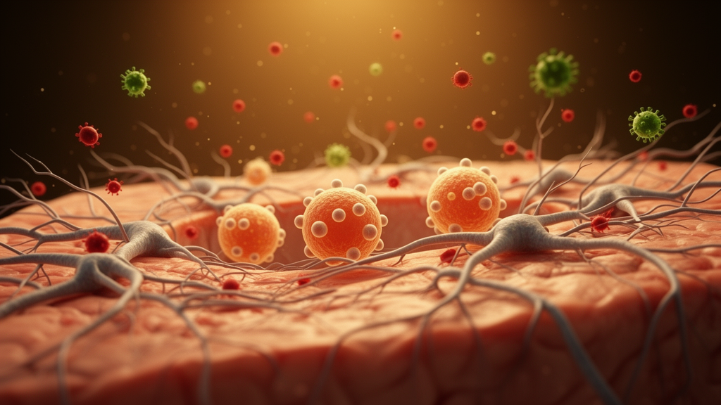
People living with HIV-1 remain vulnerable to opportunistic infections that strike the central nervous system, and few are as devastating as cryptococcal meningitis. Caused by the fungus Cryptococcus neoformans, this infection disrupts the blood-brain barrier, ignites neuroinflammation, and complicates the already delicate relationship between HIV-1 and the brain it colonizes. A new study published in PLOS Pathogens has now examined how active cryptococcal meningitis shapes the population of HIV-1 viruses circulating in the central nervous system, revealing that the interplay between virus, fungus, and host immune response is far more varied than a single model of viral evolution in the brain would suggest.
The research team, led by investigators including Maura Manion, Camille M. Lange, and Frank Maldarelli, analyzed paired samples of blood plasma and cerebrospinal fluid from thirty-nine people with HIV-1 who had active cryptococcal meningitis. All samples were collected before the participants began antiretroviral therapy, providing a snapshot of viral dynamics during a period of uncontrolled replication. Across the cohort, HIV-1 viral loads in plasma were significantly higher than those measured in cerebrospinal fluid, a pattern consistent with the blood serving as the primary reservoir of viral production. Yet the relationship between the two compartments was not uniform, and the differences that did emerge pointed to distinct mechanisms shaping viral populations behind the blood-brain barrier.
One of the clearest signals came from participants who exhibited pleocytosis, an elevated white cell count in the cerebrospinal fluid that indicates active inflammation within the central nervous system. Among these individuals, viral loads in the cerebrospinal fluid were relatively higher, and the viruses detected there more frequently used CCR5 as their coreceptor for entry into host cells. Coreceptor usage is a defining feature of HIV-1 biology: viruses that prefer CCR5 are typically associated with early infection and macrophage tropism, while those that shift toward CXCR4 tend to emerge later and target T cells more efficiently. The enrichment of CCR5-utilizing variants in inflamed cerebrospinal fluid suggests that the local environment during cryptococcal meningitis favors particular viral phenotypes, possibly because activated macrophages and microglia provide abundant CCR5-expressing targets in an inflamed brain.
To determine whether these differences reflected sustained HIV-1 replication within the central nervous system that generated genetically distinct viral lineages, a phenomenon known as viral compartmentalization, the researchers conducted a deeper genetic analysis in a subgroup of eighteen participants. Nine of these had cerebrospinal fluid HIV-1 RNA levels exceeding those in blood, while the other nine had cerebrospinal fluid levels below blood levels. The team sequenced the viral env gene, which encodes the envelope glycoprotein responsible for host cell entry, from viruses in both compartments and compared the resulting phylogenetic relationships.
Compartmentalization between cerebrospinal fluid and plasma viral populations was detected in participants from both groups, demonstrating that genetically distinct viral populations can establish themselves in the central nervous system regardless of whether cerebrospinal fluid viral loads are higher or lower than those in blood. Notably, this compartmentalization was associated with higher levels of interleukin-2 in the cerebrospinal fluid, linking the genetic separation of viral populations to the local cytokine milieu. Interleukin-2 is a signaling molecule that promotes T cell proliferation and survival, and its elevation in the cerebrospinal fluid may reflect the kind of immune activation that supports localized viral replication and evolution behind the blood-brain barrier.
The study revealed striking heterogeneity in the mechanisms that populate the central nervous system with HIV-1 during cryptococcal meningitis. In some participants, the viruses found in cerebrospinal fluid were compartmentalized from those in plasma, were exclusively macrophage-tropic, and reached concentrations exceeding 100,000 copies per milliliter of cerebrospinal fluid. This profile is consistent with sustained local replication in macrophages or microglia within the brain, producing a viral lineage adapted to that niche and largely disconnected from the circulating population. In other participants, however, the pattern suggested a different origin: HIV-1 infected cells trafficking between the plasma and cerebrospinal fluid compartments appeared to be the most likely source of the virus detected in the cerebrospinal fluid. In these cases, the central nervous system was not an independent viral sanctuary but rather a destination for infected cells migrating from the periphery.
The researchers also gained a glimpse into how these dynamics evolve after treatment begins. Post-antiretroviral therapy analysis in one participant demonstrated the emergence of distinct HIV-1 populations, indicating that the selective pressures exerted by therapy and the changing immune environment can reshape viral lineages in ways that differ between blood and brain. While the study examined this transition in only a single participant, the observation underscores that the viral populations established during active cryptococcal meningitis may have consequences that persist into the treatment era.
These findings carry significant implications for understanding HIV-1 persistence in the central nervous system and for managing neurological complications in people living with the virus. The central nervous system has long been recognized as a potential reservoir where HIV-1 can replicate independently of the blood, sheltered by the blood-brain barrier and sustained by long-lived myeloid cells such as macrophages and microglia. Cryptococcal meningitis, by compromising that barrier and driving intense inflammation, appears to create conditions that can either foster the emergence of locally adapted, macrophage-tropic viral lineages or facilitate the traffic of infected cells across compartments. The direction a given infection takes may depend on host factors such as the degree of pleocytosis and cytokine profiles, as well as on viral factors including coreceptor preference and cellular tropism.
The diversity of outcomes observed in this cohort challenges the notion that opportunistic infections of the brain act on HIV-1 in a single, predictable way. Instead, the study demonstrates that cryptococcal meningitis may have diverse effects in shaping HIV-1 viral populations, with some individuals developing compartmentalized, macrophage-tropic reservoirs in the central nervous system while others carry viruses that simply reflect ongoing exchange with the blood. As antiretroviral therapy suppresses plasma viremia in most treated individuals, understanding which patients harbor independently evolving central nervous system populations becomes increasingly important for efforts aimed at viral eradication and for explaining cases of neurological disease that persist despite effective systemic treatment. The work also highlights the value of studying HIV-1 in the context of real-world coinfections, which remain common in the regions most affected by the epidemic and may leave lasting imprints on the viral landscape that outlive the infection itself.
Subject of Research: HIV-1 compartmentalization in the central nervous system during cryptococcal meningitis
Article Title: HIV-1 compartmentalization in the central nervous system during cryptococcal meningitis: Analysis of viral, fungal, and host factors
Article References: Manion, M., Lange, C. M., Ortega-Villa, A. M., Fennessey, C. M., Keele, B. F., Moeser, M., Highbarger, H., Huik, K., Lindo, N., Gonsalves, C., Virga, J., Meya, D. B., Joseph, S. B., Boulware, D. R., Sereti, I., & Maldarelli, F. (2026). HIV-1 compartmentalization in the central nervous system during cryptococcal meningitis: Analysis of viral, fungal, and host factors. PLOS Pathogens, 22(10), e1014697. https://doi.org/10.1371/journal.ppat.1014697
Image Credits: AI Generated
DOI: 10.1371/journal.ppat.1014697
Keywords: HIV-1, cryptococcal meningitis, central nervous system, viral compartmentalization, PLOS Pathogens, macrophage-tropic HIV, blood-brain barrier, cerebrospinal fluid, compartmentalization, central, nervous, system
Cite Scienmag News
Cassandra Pierce. (October 16, 2026). Cryptococcal Meningitis Reshapes HIV-1 Populations in the Brain. Scienmag. https://scienmag.com/cryptococcal-meningitis-reshapes-hiv-1-populations-in-the-brain/
Cassandra Pierce. "Cryptococcal Meningitis Reshapes HIV-1 Populations in the Brain." Scienmag, 16 October 2026, https://scienmag.com/cryptococcal-meningitis-reshapes-hiv-1-populations-in-the-brain/. Accessed 16 October 2026.
Cassandra Pierce. "Cryptococcal Meningitis Reshapes HIV-1 Populations in the Brain." Scienmag. October 16, 2026. https://scienmag.com/cryptococcal-meningitis-reshapes-hiv-1-populations-in-the-brain/


