


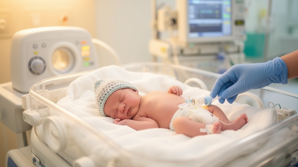
Deep at the base of the brain, in a structure barely larger than a thumb, lies the machinery that keeps every newborn alive: the brainstem. It governs breathing, heart rate, temperature regulation, feeding coordination, and the autonomic reflexes that allow a baby to transition from the womb to the outside world. For infants born months before they were meant to, the brainstem carries an extraordinary burden, and now a new study suggests that its size, measured on a routine scan, may reveal which of these fragile patients will thrive and which will struggle. Researchers at the Medical University of Vienna have produced the first set of reference values for two-dimensional brainstem measurements in extremely preterm infants, and their findings link this simple metric to cognitive and motor outcomes at two years of age.
The research, published in Pediatric Research, focused on infants born between roughly 22 and 28 weeks of gestation, the very edge of viability. Thanks to advances in neonatal intensive care, a growing number of these babies now survive, but a substantial proportion go on to face cognitive, behavioral, and motor impairments. Clinicians have long searched for early, reliable markers that could identify at-risk infants while interventions are still possible. Cerebral magnetic resonance imaging performed at term-equivalent age, the point at which a preterm infant reaches what would have been full gestational maturity, has emerged as a powerful window into the developing brain, offering far greater sensitivity than cranial ultrasound for detecting injuries in posterior and infratentorial structures, including the brainstem itself.
In the retrospective cohort study, the team analyzed infants admitted to the level IV neonatal intensive care unit at the Medical University of Vienna between August 2017 and December 2022, all of whom had undergone routine cerebral MRI before hospital discharge. From an initial pool of 315 extremely preterm infants scanned during their stay, 295 had imaging between 35 and just under 42 weeks of postmenstrual age. The median gestational age at birth was just under 26 weeks, and the mean birthweight a mere 790 grams, figures that underscore just how early this cohort entered the world. Scans were carried out on a 1.5 Tesla scanner without sedation, using a feed-and-wrap technique with a vacuum fixation mattress and ear protection so that infants could be imaged during natural sleep.
The technical heart of the study lies in its measurement protocol. Working from the midsagittal T2-weighted slice, the single midline image that cuts the brainstem lengthwise, researchers traced discrete regions of interest around each brainstem compartment. The midbrain was divided into two zones separated by the aqueduct of Sylvius, the anterior basis mesencephali and the posterior tegmentum. The pons, the prominent bulge through which vast numbers of corticocerebellar fibers pass, was outlined from its junction with the midbrain down to the pontomedullary junction. The medulla oblongata, which continues downward to the obex, was encircled along its dorsal and ventral boundaries. Reference values and z-scores were generated from 242 infants who showed no severe brain injury, meaning no high-grade intraventricular hemorrhage, no high-grade cerebellar hemorrhage, and no cystic white matter damage, and were stratified by gestational age at birth and at scanning. Repeated measurements confirmed very good intra- and interobserver reliability, with correlation coefficients of at least 0.81.
The results paint a picture of a brainstem that is both a mirror of maturity and a barometer of injury. Brainstem size increased steadily with gestational age, and infants born at 22 or 23 weeks, the threshold of viability, had the smallest measurements at term-equivalent age. More striking was the effect of neonatal brain injury. Median brainstem z-scores fell progressively with hemorrhage severity: infants without intraventricular hemorrhage scored a median of 0.13, those with low-grade bleeds 0.06, and those with high-grade hemorrhages a markedly lower negative 0.51. When the researchers grouped infants with any severe pathology, including severe hemorrhage, cerebellar bleeding, or cystic white matter injury, brainstem z-scores were significantly reduced compared with the reference cohort, with a large effect size corresponding to a Cohen’s d of 0.96. The measurements, in other words, capture global injury severity rather than isolated brainstem damage.
This pattern makes anatomical sense. The pons in particular is packed with crossing fibers that connect the cerebral cortex and cerebellum, alongside spinocerebellar and corticospinal tracts. A severe hemorrhage early in the neonatal period can unleash blood toxicity, neuroinflammation, and secondary disruption of white matter development that reverberates far beyond the site of the original bleed. A smaller brainstem at term-equivalent age may therefore reflect impaired maturation of widespread neural connectivity, a shrunk cross-section of the brain’s entire communication architecture. This interpretation helps explain why a structure that seems so remote from cognition and voluntary movement can nevertheless track developmental outcomes so closely.
And track them it does. Follow-up data at two years’ corrected age were available for about 83 percent of the cohort, assessed with the Bayley Scales of Infant and Toddler Development, third edition, using the German normative dataset and administered by certified clinical psychologists. In the reference collective of infants without severe brain injury, after adjustment for gestational age at birth and at scanning, sex, birthweight, sepsis, surgical closure of a persistent ductus arteriosus, necrotizing enterocolitis, severe retinopathy of prematurity, bronchopulmonary dysplasia, hemorrhage grade, cerebellar hemorrhage, and white matter injury, higher brainstem z-scores remained independently associated with better cognitive and motor composite scores. Each unit increase in brainstem z-score corresponded to roughly 3.6 points of cognitive score and 2.8 points of motor score. Language showed a borderline trend that narrowly missed statistical significance in this group.
When the analysis was repeated in the full cohort, including infants with severe brain injuries, the associations grew even stronger: the adjusted effects rose to about 5.1 points for cognition, 4.3 for language, and 5.7 for motor outcomes, all statistically robust. Among the four brainstem compartments, only the pons remained independently linked to cognitive and motor outcomes in the regional analysis, likely because its larger size yields more reliable measurements than the smaller midbrain and medullary segments. Meanwhile, the search for risk factors associated with a brainstem size below the tenth percentile turned up telling clues: infants with lower birthweight z-scores, higher first neonatal lactate levels, and lower first-minute APGAR scores were more likely to fall in that bottom decile, pointing to both fetal growth restriction and early hemodynamic stress as contributors. Notably, previously suspected factors such as infections and bronchopulmonary dysplasia did not reach statistical significance in this cohort.
The appeal of this biomarker lies in its simplicity. Unlike advanced quantitative techniques such as diffusion tensor imaging or myelination mapping, which demand specialized post-processing and expertise, tracing a handful of regions on a single midline slice takes minutes and uses standard radiology software available in any imaging department. That makes it an attractive candidate for routine clinical use in neonatal units that already perform term-equivalent MRI, allowing a rough but meaningful developmental risk stratification long before formal psychological testing becomes possible. If a baby’s brainstem z-score flags concern at 38 weeks, early intervention services, which are known to benefit preterm infants, could be engaged months or even years earlier than they otherwise would be.
The authors are careful to note the study’s limits. It was retrospective and single-center, so unmeasured confounders cannot be excluded, and the reference values may not apply to other gestational or imaging ranges. Two-dimensional tracings, however practical, cannot fully capture the three-dimensional complexity of brainstem anatomy, and an observational design can never prove that a small brainstem causes poor outcomes rather than simply marking shared underlying pathology. External validation in independent cohorts is required before the measurements enter routine prognostication. Still, the study opens an intriguing path forward: combining these simple morphometric traces with functional MRI, quantitative myelination measures, and neurophysiological techniques could yield multimodal, individualized forecasts of brain development. For now, the message is compelling in its economy: one of the oldest and most vital structures in the human brain, measured with a digital cursor on a single scan, may quietly reveal how the whole developing brain is destined to grow.
Subject of Research: Brainstem MRI biometry and neurodevelopmental outcomes in extremely preterm infants
Article Title: Brainstem size in extremely preterm infants: reference values and association with neurodevelopmental outcomes
Article References: Brainstem size in extremely preterm infants: reference values and association with neurodevelopmental outcomes. (n.d.). https://doi.org/10.1038/s41390-026-05529-9
Image Credits: AI Generated
DOI: 10.1038/s41390-026-05529-9
Keywords: brainstem, extremely preterm infants, cerebral MRI, term-equivalent age, intraventricular hemorrhage, neurodevelopment, Bayley-III, pons, reference values, biomarker, neonatology, white matter injury
Cite Scienmag News
Harold Sullivan. (October 15, 2026). Tiny Brainstem, Big Clues: MRI Measurements Predict Development in Extremely Preterm Infants. Scienmag. https://scienmag.com/tiny-brainstem-big-clues-mri-measurements-predict-development-in-extremely-preterm-infants/
Harold Sullivan. "Tiny Brainstem, Big Clues: MRI Measurements Predict Development in Extremely Preterm Infants." Scienmag, 15 October 2026, https://scienmag.com/tiny-brainstem-big-clues-mri-measurements-predict-development-in-extremely-preterm-infants/. Accessed 15 October 2026.
Harold Sullivan. "Tiny Brainstem, Big Clues: MRI Measurements Predict Development in Extremely Preterm Infants." Scienmag. October 15, 2026. https://scienmag.com/tiny-brainstem-big-clues-mri-measurements-predict-development-in-extremely-preterm-infants/


