


Scleroderma, known medically as systemic sclerosis, is one of the most challenging rare autoimmune diseases a physician can face. The immune system mistakenly instructs the body to produce far too much collagen, the structural protein that normally gives skin its strength and elasticity. Instead of remaining in balance, collagen accumulates relentlessly, hardening and thickening the skin and, in many patients, scarring internal organs such as the lungs, kidneys, and digestive tract. For more than half a century, the primary tool for assessing how far this fibrotic process has progressed has been remarkably low-tech: a doctor pinches the skin between thumb and forefinger and assigns a score based on how thick it feels. That tactile judgment has remained central to diagnosis, staging, and treatment monitoring since the modified Rodnan skin score became standard practice, yet it is fundamentally subjective, and two experienced clinicians can grade the same patient differently.
That subjectivity is more than an academic inconvenience. Because skin thickening is one of the most measurable manifestations of systemic sclerosis, any imprecision in tracking it ripples through every aspect of care. A patient whose disease is genuinely improving may appear stable if the examiner’s touch differs from that of the previous visit. In clinical trials, where the skin score often serves as a key outcome measure, inter-observer variability can obscure whether an experimental therapy is actually working. Researchers have long argued that the absence of a precise, objective, and repeatable way to quantify skin fibrosis is one of the major barriers slowing the development of the first approved therapies specifically targeting skin fibrosis in this disease.
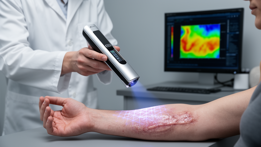
Now a team at Boston University Chobanian & Avedisian School of Medicine and the university’s Department of Biomedical Engineering reports a potential solution drawn from an unexpected corner of optics research. In a prospective cohort study published in The Lancet Rheumatology, the investigators describe the first application of a camera-based technique called spatial frequency domain imaging, or SFDI, to scleroderma. The method involves projecting patterns of harmless, non-contact light onto the skin and analyzing how those patterns are absorbed and scattered as they bounce back to a camera. Because collagen-rich, fibrotic tissue scatters and absorbs light differently from healthy skin, the returning signal carries quantitative information about what lies beneath the surface, all without ever touching the patient.
Darren Roblyer, professor of biomedical engineering and a co-author of the manuscript, noted that the technology has been a long time coming. His laboratory has spent nearly fifteen years developing SFDI for various biomedical applications, but this study marks the first time the technique has been directed at scleroderma. The adaptation matters because the optical properties that SFDI measures are sensitive to exactly the changes that define the disease: increased collagen deposition, altered tissue architecture, and the fibrotic remodeling that stiffens skin from within. In principle, a camera and a structured light source could do what a clinician’s fingers cannot—produce a number that means the same thing regardless of who takes the measurement.
To test that principle rigorously, the research team enrolled 49 people with scleroderma and 25 healthy volunteers at the Boston Medical Center Scleroderma Clinic. Each participant underwent SFDI imaging at six anatomical sites spanning the hands, forearms, and fingers, regions where skin thickening in systemic sclerosis typically appears early and progresses in characteristic patterns. The investigators then systematically compared the optical readings against the full battery of assessment tools already in clinical use. These included the physician’s touch-based skin score, ultrasound measurements of skin thickness, and a handheld device that quantifies skin hardness. Crucially, they also obtained forearm skin biopsies from 34 patients and 16 healthy volunteers, allowing the light-based measurements to be validated directly against the amount of collagen buildup visible in tissue under the microscope.
The results were strikingly consistent. Light readings were lower in people with scleroderma than in healthy individuals, and lower readings correlated with worse outcomes across every comparator: higher clinical skin scores, more scarring in the biopsy specimens, and thicker skin on ultrasound. Corresponding author Andreea Bujor, associate professor of medicine and a rheumatologist at Boston Medical Center, explained that the team compared the light readings directly with collagen accumulation in the biopsies, linking the imaging measurements to the scarring in the skin, and also tested repeatability and whether the readings changed as patients’ skin changed over time. In 11 patients, measurements were repeated longitudinally, and the optical signal tracked the direction of change in the clinical skin score, an early but important sign that the technique can follow disease evolution rather than merely snapshot it.
Perhaps the most intriguing finding was what SFDI detected in skin that doctors had scored as normal. Optical readings differed in those clinically unremarkable areas, suggesting the camera may perceive subtle collagen changes before they become palpable to even a trained hand. If confirmed in larger and longer studies, this sensitivity could shift the diagnostic timeline. Earlier detection of fibrotic change is a critical goal in systemic sclerosis because earlier diagnosis and treatment help improve prognosis and survival. A tool that flags subclinical skin involvement could allow physicians to intervene at a stage when antifibrotic strategies have a better chance of preserving tissue function, and could give trial designers a more sensitive endpoint for measuring whether new drugs slow or reverse the process.
The implications for drug development may be where the technology carries the greatest weight. With no approved therapies specifically for skin fibrosis in scleroderma, a fast, painless, non-contact measurement could help doctors track skin changes more objectively, help clinical trials detect treatment benefits more reliably, and identify subtle skin changes before they can be felt by hand, as Bujor emphasized. Clinical trials in fibrotic diseases are notoriously difficult because slow disease progression and noisy outcome measures can make even a genuinely beneficial therapy appear ineffective. An objective optical endpoint, standardized across sites and examiners, could reduce the sample sizes and trial durations needed to demonstrate efficacy, potentially lowering the cost and risk of bringing antifibrotic candidates to patients.
The study’s design also speaks to its credibility as an observational, prospective cohort effort. By anchoring the optical measurements to histological collagen content rather than relying solely on clinical scores, the researchers established a direct biological link between what the camera sees and what is actually happening in the tissue. The multi-modal comparison—touch, ultrasound, hardness, and biopsy—provides convergent validity from independent physical principles, while the repeatability testing addresses the practical question of whether the technique is stable enough for serial use in a clinic. The work received support from the National Scleroderma Foundation, the Scleroderma Clinical Trials Consortium, and the National Institutes of Health through the National Institute of Arthritis and Musculoskeletal and Skin Diseases and the National Institute of Biomedical Imaging and Bioengineering.
As with any first-in-field study, caveats remain. The cohort was modest in size, the longitudinal follow-up included only 11 patients, and the measurements were performed at a single scleroderma center, so broader validation across diverse populations and clinical settings will be needed before SFDI can join routine practice. The authors’ declared relationships with industry, disclosed as unrelated to the submitted work, underscore how much commercial and therapeutic interest is converging on fibrosis measurement. Still, the trajectory is clear: a technology refined over fifteen years in an engineering laboratory has, in a single prospective study, demonstrated that patterns of light reflected from the skin can quantify the collagen that scleroderma deposits there—objectively, painlessly, and without a single pinch. For patients whose disease has long been assessed by touch alone, the prospect of a camera that sees fibrosis coming before fingers can feel it represents a meaningful step toward earlier treatment and, ultimately, better outcomes.
Subject of Research: Spatial frequency domain imaging for objective assessment of skin fibrosis and collagen accumulation in systemic sclerosis
Article Title: BU researchers develop simple, noninvasive way to assess collagen in the skin of scleroderma patients
Article References: BU researchers develop simple, noninvasive way to assess collagen in the skin of scleroderma patients. (n.d.). Original publication
Image Credits: AI Generated
DOI: Not provided
Keywords: scleroderma, systemic sclerosis, skin fibrosis, collagen, spatial frequency domain imaging, SFDI, optical imaging, biomedical engineering, autoimmune disease, clinical trials, Boston University, The Lancet Rheumatology
Cite Scienmag News
Denise Maddox. (October 15, 2026). Camera-Based Light Imaging Offers Objective Window Into Scleroderma Skin Scarring. Scienmag. https://scienmag.com/camera-based-light-imaging-offers-objective-window-into-scleroderma-skin-scarring/
Denise Maddox. "Camera-Based Light Imaging Offers Objective Window Into Scleroderma Skin Scarring." Scienmag, 15 October 2026, https://scienmag.com/camera-based-light-imaging-offers-objective-window-into-scleroderma-skin-scarring/. Accessed 15 October 2026.
Denise Maddox. "Camera-Based Light Imaging Offers Objective Window Into Scleroderma Skin Scarring." Scienmag. October 15, 2026. https://scienmag.com/camera-based-light-imaging-offers-objective-window-into-scleroderma-skin-scarring/


