


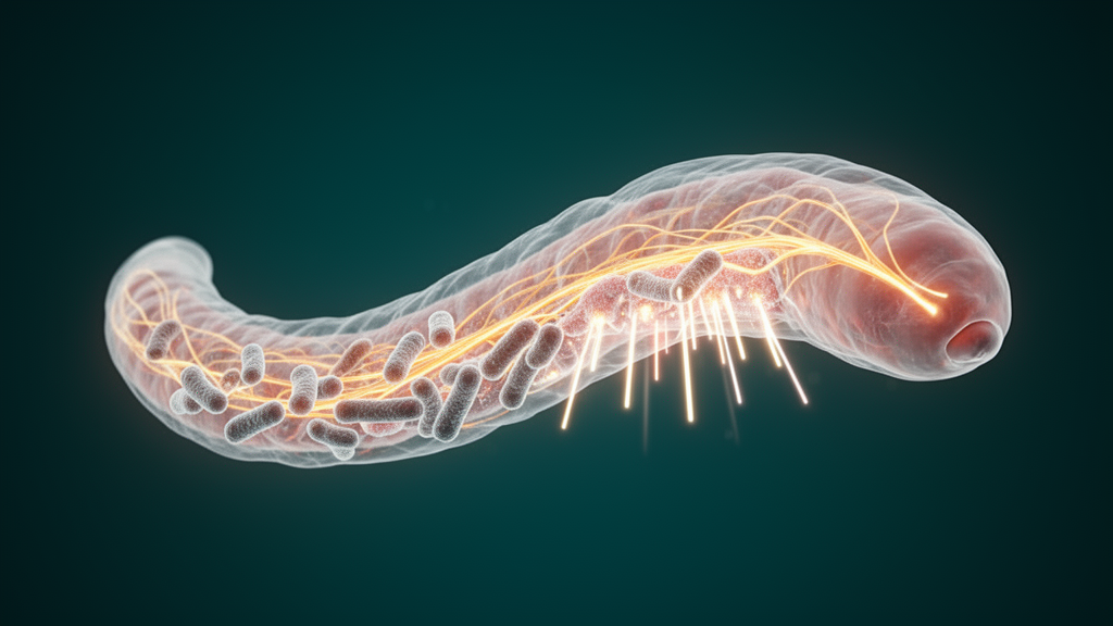
In a discovery that reshapes how scientists think about the conversation between the nervous system and the body’s outer defenses, researchers have shown that a deadly bacterium can exploit neuronal signaling to sabotage the structural integrity of an animal’s skin. Working with the roundworm Caenorhabditis elegans and the opportunistic human pathogen Pseudomonas aeruginosa, a team led by Qian Li, Yating Liu, Hanyi Chen, Weilie Xiao, and Bin Qi has mapped a signaling pathway that runs from infected neurons to the epidermis, where it corrupts the cellular recycling machinery and leaves the animal’s collagen-based cuticle in disarray. The findings, published in PLOS Biology, reveal that pathogens do not merely attack tissue directly; they can co-opt the host’s own long-range communication networks to weaken barriers from within.
The epidermis of C. elegans depends on a collagen-rich extracellular matrix, the cuticle, to serve as its first line of defense against environmental threats, including pathogenic bacteria. This structure is not static. Like bone in vertebrates, the cuticle undergoes continuous turnover, with collagen components being deposited, organized, and degraded in a carefully balanced cycle. At the heart of that degradation process sit lysosomes, the membrane-bound organelles that act as the cell’s digestive compartments. Lysosomes must maintain an acidic internal environment, achieved by proton pumps embedded in their membranes, to activate the enzymes that break down cellular cargo. When lysosomal acidification falters, maturation stalls, degradation slows, and the turnover of structural proteins such as collagen becomes erratic, undermining the precise architecture that gives the cuticle its strength.
What has remained murky is how the nervous system, which constantly monitors the environment and coordinates the animal’s behavioral and physiological responses, might influence this lysosomal housekeeping in the skin. Neurons are known to send signals that modulate immunity and stress responses across tissues, but whether pathogens could weaponize such neuro-epidermal communication to compromise the extracellular matrix had not been demonstrated. The new study set out to answer that question using the well-established infection model in which C. elegans nematodes are exposed to Pseudomonas aeruginosa strain PA14, a virulent isolate that rapidly kills the worms and has long served as a workhorse for dissecting host-pathogen interactions.
The researchers found that when worms were exposed to PA14, their neurons responded by secreting a signaling molecule the team named NSIF-1, for Neuronal Secreted Immune Factor 1. Rather than acting locally within the nervous system, NSIF-1 left the neurons and traveled to the epidermis, where it set off a cascade of damaging effects. Genetic experiments established the neuronal origin of this factor with precision: when the researchers knocked down nsif-1 expression specifically in neurons, the downstream damage to epidermal lysosomes was prevented, confirming that the signal originates in the nervous system and acts at a distance on the skin.
Inside the epidermal cells, the consequences of NSIF-1’s arrival were striking. The infected worms showed impaired lysosomal acidification, meaning the organelles failed to acidify their interiors properly. Lysosomal maturation, the process by which these compartments acquire the full complement of degradative machinery, was disrupted, and the degradation of cargo slowed accordingly. Because lysosomes govern the turnover of cuticle collagen, this failure rippled outward: the collagen structure of the cuticle became disorganized, its density dropped, and the barrier integrity of the epidermis was compromised. In practical terms, the animal’s protective armor deteriorated at precisely the moment it faced a bacterial assault, and the worms became less resistant to infection.
The molecular linchpin of this sabotage turned out to be a transcription factor called ELT-3. Transcription factors are proteins that bind to specific DNA sequences and switch target genes on or off, and ELT-3 normally plays a constructive role in the epidermis, promoting the lysosomal activity required for extracellular matrix repair. The study demonstrated that NSIF-1 interferes with ELT-3 by inhibiting its nuclear localization, that is, by preventing the protein from entering the nucleus where it would otherwise bind DNA and activate its target genes. With ELT-3 locked out of the nucleus, the transcriptional program that supports lysosomal function and collagen homeostasis went quiet, leaving the epidermis unable to maintain or repair its collagen framework.
The evidence for this mechanism came from a series of complementary genetic approaches. Worms carrying mutations in the nsif-1 gene retained healthy lysosomal function during PA14 exposure, showed enhanced collagen density in their cuticles, and survived infection at significantly higher rates than wild-type animals. These gains were not merely correlational; the restoration of lysosomal acidification and maturation in the mutants, together with the preserved organization of collagen fibers, traced a coherent causal chain from neuronal secretion to epidermal breakdown. The convergence of survival data, imaging of cuticle architecture, and molecular readouts of lysosomal state gives the pathway a level of internal consistency that strengthens the authors’ interpretation considerably.
From an evolutionary standpoint, the strategy makes grim sense for the pathogen. Pseudomonas aeruginosa is a formidable opportunistic pathogen in humans, notorious for infections of wounds, lungs in cystic fibrosis patients, and immunocompromised individuals, and it is renowned for its arsenal of virulence tactics. Subverting host signaling rather than simply mounting a brute-force assault on tissue allows the bacterium to weaken defenses with economy. Although C. elegans is a microscopic nematode, the cellular logic at play, neurons communicating with barrier tissues, lysosomes managing extracellular matrix turnover, and transcription factors coordinating repair, has deep parallels in mammalian biology. Neuro-immune crosstalk and lysosomal regulation of the extracellular matrix are active frontiers in human research, touching on fields from wound healing to fibrosis and neurodegeneration.
The identification of NSIF-1 and ELT-3 as opposing nodes in this pathway opens concrete therapeutic possibilities. If a comparable neuronal signal can be shown to act on epidermal or epithelial barriers in mammalian systems, blocking that signal, or stabilizing the activity of an ELT-3-like transcription factor, might help preserve matrix integrity during infection. Conversely, the study offers a caution: interventions that boost lysosomal activity without accounting for upstream neuronal signals may be undermined by the very pathways pathogens exploit. The authors suggest that NSIF-1 and ELT-3 represent potential targets to counteract infection-driven extracellular matrix dysregulation, a framing that invites screening for small molecules capable of restoring lysosomal acidification or protecting transcription factor localization in infected tissue.
For the broader scientific community, the work adds a vivid example to a growing catalog of pathogen strategies that operate through the host’s own regulatory circuits rather than against them. It also underscores how much a simple worm can teach. The C. elegans cuticle, with its tractable genetics and optically transparent body, allowed the team to connect a neuronal secretion, a transcription factor, an organelle, and a whole-animal outcome, survival, within a single experimental system. That end-to-end clarity is rare, and it transforms a subtle molecular observation into a complete biological narrative: a bacterium whispers to the nervous system, the nervous system unwittingly betrays the skin, and the skin’s recycling machinery grinds to a halt just when the animal needs it most. As researchers now search for echoes of this neuro-epidermal axis in higher organisms, the humble nematode has once again proven that the shortest path to understanding a complex infection may run through a millimeter-long worm.
Subject of Research: Pathogen-induced neuronal signaling that disrupts epidermal lysosomal function and collagen homeostasis in C. elegans
Article Title: Pathogen subversion of neuro-epidermal signaling impairs lysosomal function to disrupt collagen homeostasis in Caenorhabditis elegans
Article References: Li, Q., Liu, Y., Chen, H., Xiao, W., & Qi, B. (2026). Pathogen subversion of neuro-epidermal signaling impairs lysosomal function to disrupt collagen homeostasis in Caenorhabditis elegans. PLOS Biology, 24(9), e3004016. https://doi.org/10.1371/journal.pbio.3004016
Image Credits: AI Generated
DOI: 10.1371/journal.pbio.3004016
Keywords: Caenorhabditis elegans, Pseudomonas aeruginosa, NSIF-1, ELT-3, lysosomes, collagen, extracellular matrix, neuro-epidermal signaling, cuticle integrity, host-pathogen interaction, innate immunity, PLOS Biology
Cite Scienmag News
Kristina Jarvis. (October 9, 2026). How a Pathogen Hijacks Nerve Signals to Break Down a Worm’s Skin. Scienmag. https://scienmag.com/how-a-pathogen-hijacks-nerve-signals-to-break-down-a-worms-skin/
Kristina Jarvis. "How a Pathogen Hijacks Nerve Signals to Break Down a Worm’s Skin." Scienmag, 9 October 2026, https://scienmag.com/how-a-pathogen-hijacks-nerve-signals-to-break-down-a-worms-skin/. Accessed 9 October 2026.
Kristina Jarvis. "How a Pathogen Hijacks Nerve Signals to Break Down a Worm’s Skin." Scienmag. October 9, 2026. https://scienmag.com/how-a-pathogen-hijacks-nerve-signals-to-break-down-a-worms-skin/


