


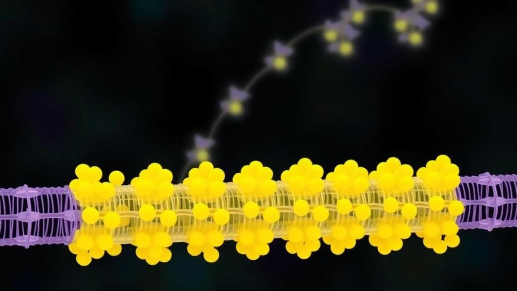
Human cells manufacture thousands of integral membrane proteins, from transporters and channels to receptors, and nearly all of them are threaded into the endoplasmic reticulum membrane while they are still being synthesized by ribosomes. For decades, the central player in this process was thought to be Sec61, the protein-conducting channel that docks onto the ribosome and can either push a nascent chain across the lipid bilayer or release it laterally into the membrane. But work over the past several years has overturned the simple picture. Potent inhibitors that plug the lateral gate of Sec61 revealed that many, and possibly most, multipass membrane proteins are inserted by alternative routes, facilitated by members of the universally conserved Oxa1 superfamily of membrane insertases. A new study published in Nature Structural & Molecular Biology now provides the most detailed view yet of how two of these insertase systems, the ER membrane protein complex (EMC) and the multipass translocon (MPT), cooperate at the ribosome to build complex membrane proteins.
The research team, led by Robert Keenan at the University of Chicago together with Ramanujan Hegde at the MRC Laboratory of Molecular Biology and Zhe Ji at the Shanghai Institute of Biochemistry and Cell Biology, combined three complementary techniques: selective ribosome profiling, cryo-electron tomography and site-specific photocrosslinking. Each method answers a different question. Ribosome profiling reveals which proteins are being synthesized on ribosomes engaged with a particular factor and at what point in the sequence. Cryo-electron tomography shows the three-dimensional architecture of the assembly in native membranes. Photocrosslinking maps the physical contacts between the nascent chain and its molecular chaperones. Together, these approaches allowed the researchers to determine the substrate range, the timing and the mechanism of EMC recruitment during multipass protein biogenesis in human cells.
The first surprise came from the profiling data. The researchers affinity-purified ribosome complexes associated with tagged EMC subunits from human embryonic kidney cells and sequenced the ribosome-protected messenger RNA fragments. Of roughly 5,800 proteins detected, multipass membrane proteins were the most enriched class, but EMC recovered only a subset of them, and enrichment increased with the number of transmembrane domains. This contrasted with the multipass translocon, which had previously been shown to engage nearly all multipass proteins regardless of their size. Comparing the two datasets directly, the team found that about half of multipass transcripts were enriched by both EMC and MPT, about one-third by MPT alone, and essentially none by EMC alone. The conclusion was inescapable: EMC is recruited to ribosomes that are already bound to the multipass translocon, arriving later in the synthesis of a growing membrane protein.
The timing of EMC arrival proved to be remarkably informative. The multipass translocon, composed of the GEL, PAT and BOS complexes assembled around Sec61, features a central membrane cavity that can accommodate roughly six to eight transmembrane domains, along with a small cytosolic vestibule that can shield about 60 to 90 amino acid residues. The researchers found that EMC typically arrived at the translocon when between two and eight transmembrane domains had emerged from the ribosome, and that the exact moment depended on the properties of the substrate. Proteins with long cytosolic segments early in their sequence recruited EMC sooner, while proteins with short cytosolic loops waited until more transmembrane domains had accumulated. The midpoint of EMC onset shifted from about five transmembrane domains for proteins with few cytosolic residues to just over two domains for proteins with more than 120 cytosolic residues in their first two cytosolic segments.
These observations led the team to a simple but powerful model: EMC is recruited when the growing nascent chain begins to exceed either the cytosolic or the intramembrane capacity of the multipass translocon. Once the central cavity is full of transmembrane domains, or the cytosolic vestibule can no longer shield the exposed hydrophobic and charged segments, the substrate begins to spill out, and EMC is there to catch it. Crucially, once EMC engaged, it stayed bound until synthesis was complete, even when the multipass translocon itself repeatedly disengaged and re-engaged during the synthesis of long cytosolic or lumenal segments. This stability distinguishes EMC from the more dynamic MPT and OST-A components, whose association with the ribosome can be reversibly disrupted as the substrate’s topology demands.
To see the assembly directly, the researchers programmed human microsomes with a stalled four-transmembrane-domain intermediate of ABCD4, a six-transmembrane ABC transporter, and imaged them by cryo-electron tomography. Among the ribosome-bound particles, they resolved two classes of multipass translocon: one containing Sec61, GEL, PAT and BOS, and a second that carried an additional elongated density spanning the membrane near Sec61 and PAT. That density was EMC, which could be unambiguously fitted as a single rigid body. The structure revealed a wedge-shaped cytosolic domain of EMC packing against the ribosomal RNA and the ribosomal protein eL31, creating a large semi-enclosed cytosolic chamber flanked by the ribosome, EMC and the PAT complex. This chamber is several-fold larger and more exposed than the MPT cytosolic vestibule, providing space for substrates that can no longer be accommodated within the multipass translocon itself.
The membrane-embedded heart of the structure was equally revealing. The conserved hydrophilic groove formed by the Oxa1 insertase module of EMC, comprising the subunits EMC3 and EMC6 and possibly gated by EMC4, faces the open end of the multipass translocon central cavity. This arrangement defines an expanded lipid-filled cavity bounded by EMC, MPT and Sec61. Notably, the hydrophilic grooves of GEL and EMC sit on opposite sides of the assembly, roughly 40 and 100 angstroms from the ribosome exit tunnel respectively, meaning that transmembrane domains emerging from the ribosome have at least two potential routes into the membrane. On the opposite face of EMC lies the holdase surface previously shown to engage an assembly intermediate of the voltage-gated calcium channel Cav1.2, and superimposition showed that this binding mode is fully compatible with EMC’s position at the translocon. EMC thus sits at a crossroads, interfacing cotranslationally with nascent chains emerging from the ribosome on one side and posttranslationally with inserted but unassembled substrates on the other.
The photocrosslinking experiments confirmed this picture at residue-level resolution. In a two-transmembrane intermediate of ABCD4, probes placed in the first two transmembrane domains crosslinked to TMCO1 of the GEL complex and to Sec61α, showing that the newly inserted pair resides within the multipass translocon central cavity. In a four-transmembrane intermediate, those same probes instead crosslinked to EMC3, EMC4, EMC7 and EMC10, while the newly inserted third and fourth transmembrane domains now crosslinked to GEL. In other words, as the chain grew, the earlier transmembrane pair migrated from the GEL side of the cavity to the insertase side of EMC, while the newer pair took its place near GEL. The crosslinking patterns followed periodicities consistent with alpha-helical substrates in favored orientations, indicating that both EMC and GEL directly engage the transmembrane domains and shield their most hydrophilic faces. This defines the insertase side of EMC as a cotranslational chaperone, a function that parallels observations of the bacterial Oxa1 superfamily member YidC, whose depletion perturbs the folding but not the topology of the multipass transporter LacY.
The study resolves several long-standing questions about how the cell triages its membrane protein traffic. Because MPT binds preferentially to closed Sec61 channels and is therefore recruited for nearly all multipass proteins, while EMC is recruited by the exposure of already-inserted transmembrane domains, the substrate ranges of the two systems overlap only partially. Proteins whose transmembrane domains remain sequestered within the multipass translocon cavity throughout synthesis never robustly recruit EMC. The partial redundancy of EMC and GEL as insertases is likewise explained: GEL, positioned closer to the ribosome exit tunnel, likely has priority access, with EMC inserting transmembrane pairs that GEL skips. This may also explain why some organisms lack GEL altogether, compensating with functionally analogous proteins or with EMC acting alone at membrane-bound ribosomes.
What emerges is a modular, substrate-responsive factory in which Sec61, the multipass translocon and EMC assemble and reassemble according to the topological demands of each nascent chain. EMC’s multiple activities as an insertase, a transmembrane-domain chaperone and a holdase for assembly intermediates allow it to accommodate the widely varying biophysical needs of the roughly 2,500 multipass membrane proteins encoded in the human genome. Given that defects in membrane protein biogenesis underlie numerous diseases, from channelopathies to transporter deficiencies, understanding how these molecular machines cooperate opens the door to strategies for correcting misfolded or mistargeted membrane proteins. The EMC-bound translocon, once an invisible intermediate, is now a defined structural entity, and its architecture provides a framework for dissecting the remaining rules of membrane protein assembly in health and disease.
Subject of Research: Cotranslational biogenesis of multipass membrane proteins by the EMC-bound translocon at the endoplasmic reticulum
Article Title: Cotranslational membrane protein biogenesis by an EMC-bound translocon
Article References: Rollins, M. G., Tang, J., Wan, Y., Sundaram, A., Wu, H., Li, Q., Fedry, J., Hegde, R. S., Ji, Z., & Keenan, R. J. (2026). Cotranslational membrane protein biogenesis by an EMC-bound translocon. Nature Structural & Molecular Biology. https://doi.org/10.1038/s41594-026-01889-2
Image Credits: AI Generated
DOI: 10.1038/s41594-026-01889-2
Keywords: EMC, multipass translocon, Sec61, endoplasmic reticulum, membrane protein biogenesis, ribosome profiling, cryo-electron tomography, photocrosslinking, Oxa1 superfamily, transmembrane domains, protein insertion, molecular chaperone
Cite Scienmag News
Drew Townsend. (October 8, 2026). How the cell builds its membrane proteins: new view of the EMC-bound translocon. Scienmag. https://scienmag.com/how-the-cell-builds-its-membrane-proteins-new-view-of-the-emc-bound-translocon/
Drew Townsend. "How the cell builds its membrane proteins: new view of the EMC-bound translocon." Scienmag, 8 October 2026, https://scienmag.com/how-the-cell-builds-its-membrane-proteins-new-view-of-the-emc-bound-translocon/. Accessed 8 October 2026.
Drew Townsend. "How the cell builds its membrane proteins: new view of the EMC-bound translocon." Scienmag. October 8, 2026. https://scienmag.com/how-the-cell-builds-its-membrane-proteins-new-view-of-the-emc-bound-translocon/


