


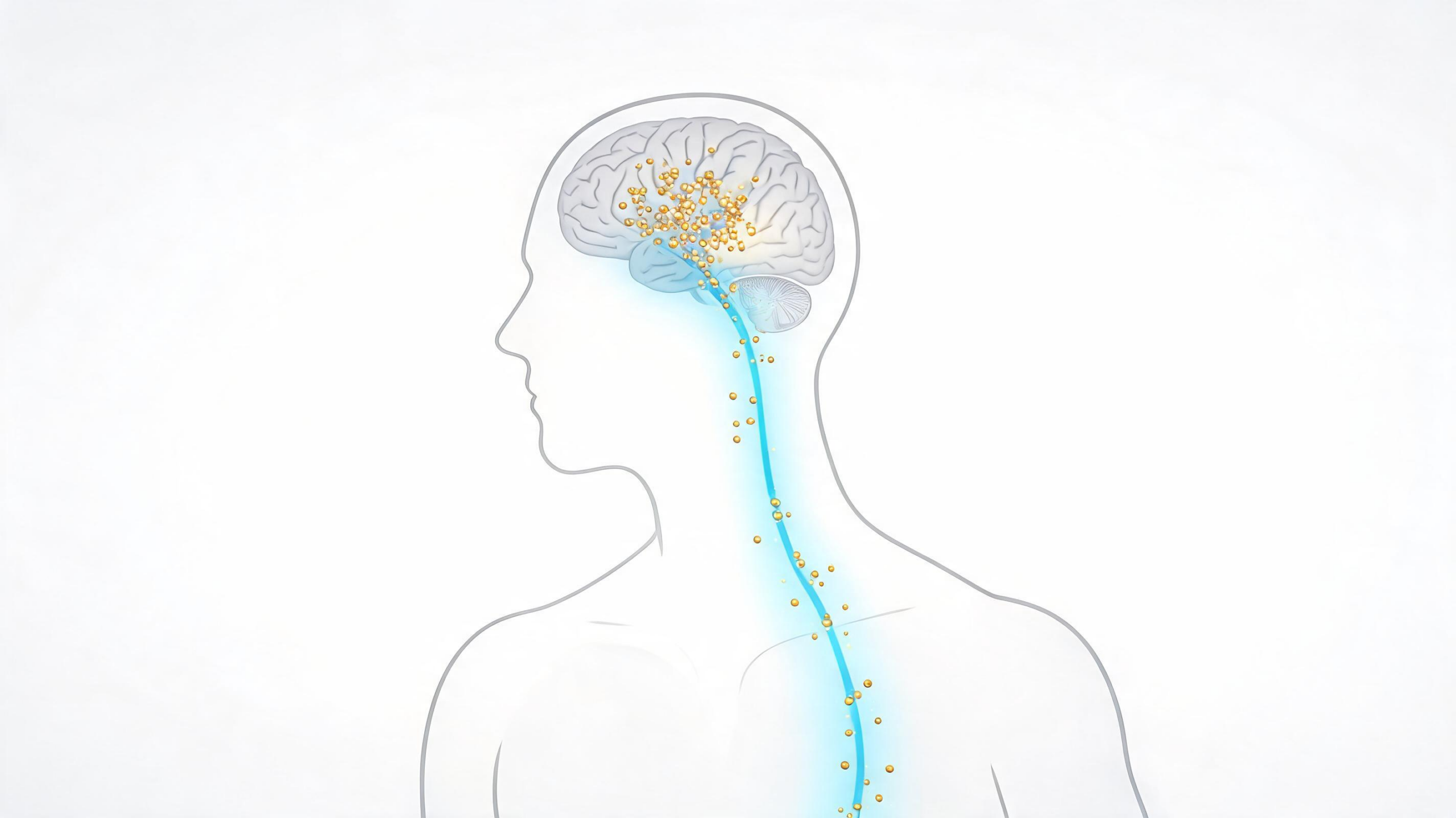
Melatonin has long been celebrated as the brain’s chemical messenger of darkness, the hormone that rises at nightfall and gently steers the body’s circadian machinery. Yet a growing body of research has revealed that this indolamine, formally known as N-acetyl-5-methoxytryptamine, is far more than a sleep signal. It is produced not only by the pineal gland but also by mitochondria in peripheral cells, and it accumulates in tissues such as the liver, kidney, heart, placenta, and pancreas. It scavenges reactive oxygen species, dampens inflammation, helps regulate blood pressure, and has been implicated in counteracting the beta-amyloid accumulation associated with Alzheimer’s disease. Reduced endogenous melatonin levels have been linked to neurodegenerative conditions including Alzheimer’s, Parkinson’s disease, dementia, and schizophrenia. The trouble, until now, has been that actually measuring melatonin inside these extrapineal tissues has required bulky, expensive laboratory instrumentation and painstaking sample preparation.
A research team led by Marcos Vilas Boas Filho and Valber de Albuquerque Pedrosa at São Paulo State University (UNESP) in Botucatu, Brazil, working with colleagues at the same institution, has now demonstrated a compact alternative: an electrochemical immunosensor built on a bioconjugated gold electrode that can generate melatonin-associated signals directly in homogenized kidney, liver, and heart tissue from laboratory rats. Published in Discover Electrochemistry, the study is billed as the first proof-of-concept demonstration of electrochemical immunosensing for melatonin in extrapineal tissue. Rather than seeking the lowest detection limit in the field, the team set out to show that antibody-based molecular recognition could be married to electrochemical transduction in matrices as chemically hostile as tissue homogenates.
The analytical chemistry underlying conventional melatonin measurement is well established. Techniques such as chemiluminescence, fluorometry, ultraviolet–visible spectrophotometry, gas chromatography–mass spectrometry, and high-performance liquid chromatography all deliver robust performance, but they demand multiple instrumental platforms, labor-intensive preparation, and long analysis times. Melatonin’s intrinsic photoreactivity compounds the difficulty, requiring manipulation under light-restricted conditions to prevent degradation. Electrochemical biosensors have emerged as attractive alternatives because of their low detection limits, operational simplicity, and rapid response, and recent years have seen nanostructured platforms achieve impressively low limits of detection in serum, urine, food, and pharmaceutical samples. A paper-based graphite electrode, a molecularly imprinted polymer platform, and a sensor incorporating core–shell Cu@Pt nanoparticles have all reported submicromolar detection. But nearly all of these rely on the direct electrochemical oxidation of melatonin, an approach vulnerable to electrode fouling, matrix interference, and overlapping signals from other electroactive compounds—and none had been applied to extrapineal tissue.
The Brazilian team’s strategy inverts that logic. Instead of oxidizing melatonin directly, they built an indirect sensing architecture in which the hormone is captured by an immobilized antibody, and its presence is read out as a measurable suppression of a redox probe’s current. The fabrication begins with a gold electrode 1.7 millimeters in diameter, onto which a self-assembled monolayer of 11-mercaptoundecanoic acid is formed by gold–sulfur bonding, exposing terminal carboxyl groups. These groups are then activated with the classic EDC/NHS coupling chemistry, generating reactive NHS-esters that covalently link to free amine groups on a polyclonal anti-melatonin antibody during overnight incubation at 4 degrees Celsius. The result is the Au/SAM-MUA/anti-ME interface: a stable, antibody-decorated surface in which every subsequent molecular event translates into an electrical signature.
Characterization of the assembly followed the standard toolbox of electroanalytical science. Cyclic voltammetry using the ferricyanide/ferrocyanide couple as a redox probe showed a progressive decline in peak current as each layer was added, confirming that the growing protein and organic films were hindering electron transfer as designed. Electrochemical impedance spectroscopy told the same story quantitatively: the charge-transfer resistance of the bare gold electrode stood at just 5 kilo-ohms, rising to 13 kilo-ohms after monolayer formation, 29 kilo-ohms after EDC/NHS activation, 30 kilo-ohms after antibody immobilization, and a marked 40 kilo-ohms once melatonin bound to the antibody layer. That final jump, the team notes, is the analytical heart of the device—each melatonin molecule captured at the surface adds insulating mass, physically blocking diffusion of the redox probe and deepening the measurable signal.
With square-wave voltammetry optimized at a frequency of 100 hertz, a step potential of 5 millivolts, and a pulse amplitude of 20 millivolts, the researchers calibrated the sensor against commercial melatonin standards across a linear range of 20 to 120 micromolar. The calibration curve carried a negative slope of –0.0034, exactly what the suppression mechanism predicts: the blank current of 0.59 microamperes fell to 0.18 microamperes at 120 micromolar melatonin. The derived figures of merit were a limit of detection of approximately 4 micromolar, a limit of quantification of 14 micromolar, and a striking electrochemical sensitivity of 250 microamperes per micromolar per square centimeter—among the highest sensitivities reported for any electrochemical melatonin platform, and the second highest overall. Recovery analysis with spiked samples reached 99.8 percent, and the sensor held 96.3 percent of its signal between consecutive measurement days, retaining functional integrity for up to eight days before the biological layer required re-immobilization.
Selectivity testing against common biological interferents revealed both strengths and honest limits. Serotonin, dopamine, and uric acid each shifted the redox signal by only 0.8 to 4.0 percent, well within acceptable tolerances. But ascorbic acid alone produced a 9.5 percent suppression, and a mixture of all interferents together caused an 11 percent deviation—statistically significant and a reminder that nonspecific matrix effects can creep into any antibody-based measurement in complex fluids. The authors attribute this partly to possible conformational changes in the antibody at certain pH values, which may partially expose the underlying electrode surface to blocking by other molecules. They are careful to frame the sensor’s selectivity as demonstrable but not yet definitive under all biological conditions.
The biological application was where the platform earned its novelty claim. Thirty male Wistar rats were divided into a treated group receiving intraperitoneal melatonin at 25 milligrams per kilogram three times weekly for four weeks, and a control group receiving saline. Liver, heart, and kidney samples were harvested, homogenized, and spiked with a known melatonin standard before analysis. Across all three tissues, successive additions of homogenate produced the characteristic progressive suppression of the ferri/ferrocyanide anodic current, and tissues from melatonin-treated animals consistently generated stronger current suppression than control samples. Kidney homogenates produced the greatest effect, followed by heart and liver—a pattern the researchers note aligns with known physiology, since the kidney is central to eliminating melatonin metabolites, the liver metabolizes the hormone via cytochrome P450 enzymes, and cardiac tissue harbors extrapineal melatonin and receptors tied to cardiovascular regulation.
The team is appropriately measured about what the tissue data mean. Because the current-response plots in the homogenates lacked sufficient linearity to derive formal detection limits for those matrices, and because no direct comparison with HPLC or LC–MS/MS was performed, the tissue signals are presented strictly as preliminary, qualitative proof-of-concept responses rather than precise quantifications. Still, the implications are considerable. The work establishes, for the first time, that an antibody-functionalized electrochemical interface can register melatonin-associated differences in kidney, liver, and heart tissue—opening a path toward rapid, point-of-care monitoring of hormone distribution in contexts where chromatography is impractical. The researchers say future work will focus on validating the platform against established chromatographic methods and implementing matrix-matched calibration to sharpen quantitative accuracy, potentially extending the technology to studies of circadian biology, neurodegenerative disease research, and antioxidant therapy monitoring where melatonin’s reach beyond the brain matters most.
The choice of an indirect immunosensing format carries practical implications worth underscoring. Because melatonin itself is not oxidized at the electrode surface, the many electroactive species that populate tissue homogenates—ascorbate, urate, catecholamines—compete far less directly for the analytical signal. The trade-off is kinetic and structural: antibody–antigen binding is slower than a simple electron-transfer event, and the biological recognition layer is inherently fragile, which is why the team found the interface required re-immobilization after roughly eight days of use. Such operational lifetimes are typical of protein-based sensors and represent a genuine engineering constraint for any future field deployment.
The tissue-specific response pattern observed in the rat study also merits interpretation. The strongest suppression in kidney homogenates is consistent with the organ’s role as the principal route of melatonin metabolite excretion, while the hepatic signal reflects cytochrome P450-mediated metabolism, the dominant catabolic pathway for the hormone in mammals. Cardiac tissue, meanwhile, is of particular interest because melatonin receptors expressed in myocardium have been linked to blood pressure regulation and cardioprotection, making a rapid tissue-level assay potentially valuable in cardiovascular research.
Methodologically, the spiking approach used in the proof-of-concept experiments deserves note. By adding a known commercial melatonin standard to each homogenate, the researchers could verify that the antibody layer remained functional even amid the proteins, lipids, and salts of a crude tissue matrix. The absence of a chromatographic cross-check, however, means the absolute endogenous concentrations in treated versus control animals remain unknown. Establishing that correlation, alongside matrix-matched calibration curves, will be the decisive next step in determining whether this bioconjugated gold interface can evolve from a qualitative indicator of melatonin-associated tissue responses into a genuinely quantitative analytical instrument for circadian and biomedical research.
Subject of Research: Development of a bioconjugated gold electrochemical immunosensor for detecting melatonin in extrapineal rat tissues
Article Title: Electrochemical melatonin detection in extrapineal tissue using a bioconjugated sensor
Article References: Filho, M. V. B., Agneis, M. L. G., de Souza, M. C., Gavioli, V. D., de Castro, G. R., Seiva, F. R. F., de Almeida Chuffa, L. G., & de Albuquerque Pedrosa, V. (2026). Electrochemical melatonin detection in extrapineal tissue using a bioconjugated sensor. Discover Electrochemistry, 3(1), Article 75. https://doi.org/10.1007/s44373-026-00162-x
Image Credits: AI Generated
DOI: 10.1007/s44373-026-00162-x
Keywords: melatonin, immunosensor, electrochemistry, biosensor, gold electrode, extrapineal tissue, square-wave voltammetry, electrochemical impedance spectroscopy, Wistar rats, circadian rhythm, antioxidant, tissue homogenate
Cite Scienmag News
Cassandra Pierce. (September 10, 2026). Bioconjugated Gold Sensor Tracks Melatonin Beyond the Brain. Scienmag. https://scienmag.com/bioconjugated-gold-sensor-tracks-melatonin-beyond-the-brain/
Cassandra Pierce. "Bioconjugated Gold Sensor Tracks Melatonin Beyond the Brain." Scienmag, 10 September 2026, https://scienmag.com/bioconjugated-gold-sensor-tracks-melatonin-beyond-the-brain/. Accessed 10 September 2026.
Cassandra Pierce. "Bioconjugated Gold Sensor Tracks Melatonin Beyond the Brain." Scienmag. September 10, 2026. https://scienmag.com/bioconjugated-gold-sensor-tracks-melatonin-beyond-the-brain/


