

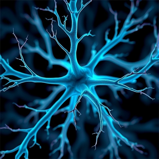
Why would a tiny tissue sample from a rat’s knee hold the key to unlocking the mysteries of chronic jaw pain? The question is not as bizarre as it sounds. Temporomandibular disorders, a group of more than 30 conditions causing pain and dysfunction in the jaw joint, affect millions of people worldwide, yet for the vast majority of cases the underlying cause remains maddeningly obscure. A new study from the University of Pittsburgh, published in npj Imaging, has taken a critical step toward changing that by deploying a custom-built imaging pipeline capable of mapping the three-dimensional architecture of nerves inside dense joint tissue with unprecedented clarity. And the first tissue to yield its secrets is, indeed, from the rat knee.
The temporomandibular joint, or TMJ, acts as a sliding hinge connecting the jawbone to the skull, making it essential for talking, chewing, and yawning. When something goes wrong with the muscles or the joint itself, the resulting pain can be debilitating, yet clinicians are often baffled because standard radiographs of patients with and without severe pain can look remarkably similar. Alejandro Almarza, professor of oral and craniofacial sciences at Pitt’s School of Dental Medicine and the study’s senior author, has long suspected that the answer lies not in bone structure but in the density, branching patterns, and molecular identity of the nerves buried deep within the joint. Visualizing those nerves, however, has been nearly impossible with conventional techniques. Histology, the traditional method of slicing tissue into thin sections and staining them, obliterates the three-dimensional context, leaving researchers blind to how neural networks actually weave through cartilage, bone, and ligament.
To overcome this limitation, Almarza turned to two colleagues at Pitt’s Center for Biologic Imaging: Simon Watkins, a distinguished professor of cell biology who has spent decades building some of the world’s most advanced custom light-sheet fluorescence microscopes, and Alan Watson, an associate professor whose expertise in tissue clearing protocols and high-performance computing provides the other half of the equation. Together, the trio set out to adapt and refine a technique called tissue clearing, which renders an entire intact piece of tissue optically transparent so that fluorescently labeled structures inside can be imaged in three dimensions. Light-sheet microscopy accelerates this process dramatically by sweeping a thin plane of laser light through the cleared tissue, capturing near-confocal resolution images at speeds that would be unthinkable with point-scanning methods, all while causing minimal photodamage.
The team systematically compared two clearing methods on rat knee joint samples, a stand-in chosen because the knee, like the TMJ, is a heavily innervated synovial joint. The first, a previously established protocol called PEGASOS designed for bone-containing tissues, left behind stubborn autofluorescent proteins that blocked laser penetration and generated high background noise that obscured fine nerve fibers. The second method, dubbed c-Clear and developed in-house at the CBI, introduced a critical innovation: a 24-hour photobleaching step prior to staining. By inactivating those autofluorescent molecules before introducing fluorescently tagged antibodies that bind specifically to neurofilament, a structural protein abundant in nerve cells, c-Clear allowed the antibodies to label the entire neural network without interference, producing a crisp, three-dimensional wiring diagram of the joint’s innervation.
The resulting image quality is as breathtaking as it is data-intensive. A single three-dimensional nerve map of one rat knee consumes roughly one terabyte of information, and the full project dataset balloons to approximately 16 terabytes. Handling such a deluge requires the CBI’s formidable computational backbone: seven petabytes of total storage and a dedicated H200 GPU cluster used for stitching, cleaning, and analyzing every volume. Watson noted that these high-speed imaging modalities generate data on a scale that commercial solutions simply cannot handle, which is why the center has built its own parallelized processing pipelines from scratch. The entire workflow, from the start of clearing to the final reconstructed image, takes between six and eight weeks per sample, a testament to both the labor involved and the unprecedented level of detail it yields.
The significance of this advance extends far beyond a single rodent knee. The work was conducted as part of the ReJoin Consortium, a $50 million project funded through the National Institutes of Health’s HEAL Initiative that aims to map nerve architecture across multiple joints, species, and disease states to decode how neural wiring relates to pain signaling. With c-Clear now validated on some of the most optically challenging tissue the consortium has yet tackled, Almarza is finally turning his attention to the structure he set out to study all along: the human temporomandibular joint. The foundational datasets, believed to be the first of their kind, have been made publicly available through the NIH’s SPARC Portal so that researchers worldwide can interrogate them.
Looking ahead, the team faces the daunting task of quantification. Producing stunning images is one thing; extracting meaningful biological metrics, such as nerve fiber density, branch angles, or spatial relationships with specific cell types, demands entirely new computational pipelines. Almarza and his collaborators are now building machine-learning tools capable of segmenting and tracing individual nerve fibers across terabytes of voxel data, a challenge that sits at the frontier of both neuroscience and image analysis. The ultimate goal is to identify objective structural signatures that distinguish painful joints from pain-free ones, something that could one day guide targeted treatments or even preventive interventions.
For the millions of people who suffer from temporomandibular disorders without a clear diagnosis, the research offers a tangible glimmer of hope. If scientists can pinpoint exactly how the nerve maps in a painful TMJ differ from those in a joint that appears identical on an X-ray yet causes no discomfort, the door opens to therapies that address the true source of the pain rather than merely managing symptoms. In a field that has long been forced to rely on subjective patient reports and ambiguous imaging, the ability to visualize the complete neural landscape of a joint in three dimensions represents nothing short of a paradigm shift, and it all began with a rat’s knee.
Subject of Research: Animal tissue samples (rat knee joints, with implications for temporomandibular joint nerve architecture)
Article Title: Advanced tissue clearing and three-dimensional imaging approaches to visualize neural innervation in the rat knee joints
News Publication Date: 13 May 2026
Web References: https://www.nature.com/articles/s44303-026-00167-6; https://sparc.science/datasets/673
References: DOI: 10.1038/s44303-026-00167-6
Image Credits: Aimee Obidzinski, University of Pittsburgh
Keywords: tissue clearing, light-sheet fluorescence microscopy, temporomandibular joint, neural innervation, pain signaling, high-resolution imaging, c-Clear, PEGASOS, ReJoin Consortium, HEAL Initiative, three-dimensional imaging, bioengineering, cell biology, dentistry


