


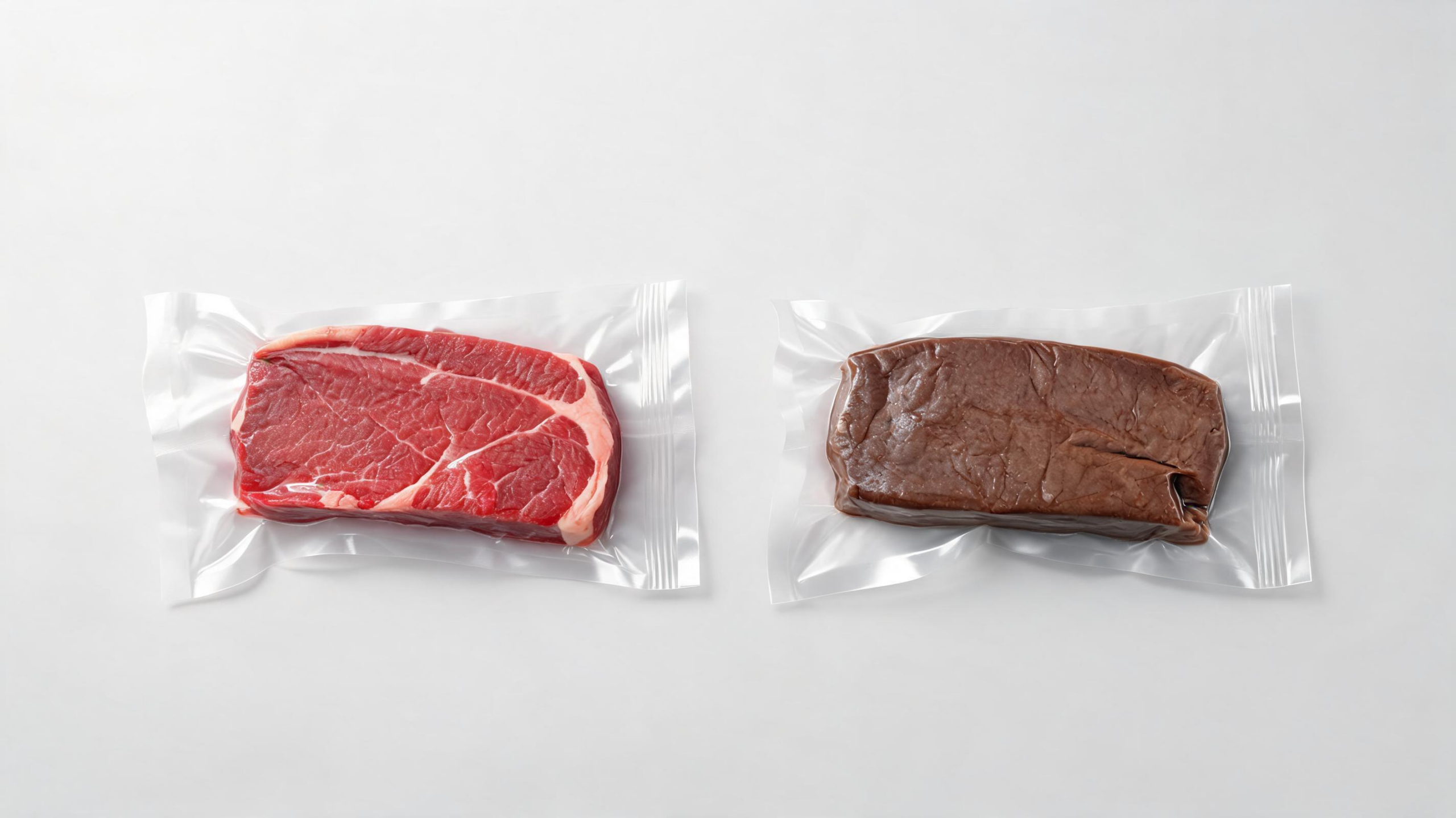
Vacuum packaging is supposed to protect beef from oxygen, extend its useful life and create the conditions for wet aging. Yet in German slaughterhouses, some beef develops an irreversible gray or brown discoloration within the first days after packaging, even when the meat appears to have been handled under normal conditions. A new investigation has examined whether the explanation lies inside the muscle itself—in its antioxidants, energy-related molecules, acidity or ability to control oxidation. The study found clear chemical signatures of discoloration, but no single intrinsic factor that could be identified as its cause. Instead, the findings point toward a complicated interaction between muscle chemistry and events occurring during slaughter, chilling, cutting and packaging.
The issue matters because meat color strongly influences whether consumers accept a product. Beef stored without oxygen commonly appears dark red, purple or violet because deoxymyoglobin dominates in the sealed package. That appearance can change when the package is opened and the surface meets air, allowing oxymyoglobin to form and produce a familiar bright red color. The phenomenon examined in this study is different. The affected samples became lighter, less red and more yellow, with gray or brown areas that did not readily recover. Such discoloration can lead consumers to reject the meat, creating economic losses and contributing to food waste even when color alone does not establish that the product is unsafe.
Color in beef is governed largely by myoglobin, the muscle protein that binds oxygen. Myoglobin can exist in several redox states: deoxymyoglobin, in which iron remains in the reduced Fe2+ state without oxygen; oxymyoglobin, in which oxygen is bound and the meat appears red; and metmyoglobin, in which the iron has been oxidized to Fe3+ and the meat tends to look brown. Muscle contains systems that can reduce metmyoglobin and restore the reduced forms, but those systems weaken after slaughter as cellular metabolism changes. Lipids and other proteins can also oxidize, generating reactive compounds that may promote further myoglobin oxidation. The researchers therefore tested a broad set of measurements rather than searching only for one antioxidant or one packaging defect.
Over four months, the team screened a commercial wet-aging facility on 30 different days. They collected nine visibly discolored samples and nine reference samples from 18 animals. The material came from beef hind muscles commonly vacuum-packaged as a single piece for aging and distribution, including the gluteobiceps, tensor fasciae latae and vastus lateralis. Samples were identified five to eight days after slaughter, frozen at minus 18 degrees Celsius and transported for laboratory analysis. Because the discoloration was rare, appeared after packaging and could not be reproduced reliably in the laboratory, sampling had to take place during routine commercial operations. The reference pieces came from the same groups of animals and handling conditions, although the researchers could not always trace each individual piece to the same animal.
The team first quantified color using the CIELAB system, in which L* represents lightness, a* the green-to-red axis and b* the blue-to-yellow axis. Compared with the controls, discolored beef had an average L* value of 43.21 rather than 38.63, an a* value of 9.42 rather than 11.21 and a b* value of 12.48 rather than 10.39. In practical terms, the affected meat was lighter, less red and more yellow. Its redness index, calculated as a/b, fell from 1.08 in controls to 0.75 in discolored samples. Reflectance spectra were also used to estimate the relative abundance of myoglobin redox states. Discolored samples contained less deoxymyoglobin and oxymyoglobin but more metmyoglobin: 1.13, 2.02 and 1.01, respectively, compared with 1.30, 2.22 and 0.79 in controls.
Those measurements confirmed that the visual change was consistent with increased myoglobin oxidation, but they did not reveal what initiated it. The researchers measured pH, redox potential, total reducing ability, lactate, the metabolites NAD+ and NADH, and two antioxidant compounds, alpha-tocopherol and beta-carotene. Almost all of these variables failed to differ significantly between the groups. The average pH was nearly identical—5.60 in controls and 5.61 in discolored beef—placing both within the typical ultimate pH range for beef. Redox potential was 457.00 millivolts in controls and 459.87 millivolts in affected samples, while total reducing ability was 4.07 and 4.12, respectively. These results did not support a major difference in overall oxidation potential or nonspecific reducing capacity.
The only non-color-related measurement that showed a statistically significant difference was lactate. Control samples contained 3.90 milligrams per gram, or about 43.8 millimoles per kilogram, whereas discolored samples contained 5.61 milligrams per gram, or about 63.0 millimoles per kilogram. Even that result does not demonstrate causation. Lactate is generally associated with color stability because it can support glycolytic activity and, under some conditions, increase the production of NADH, a reducing equivalent used by enzyme systems that convert metmyoglobin back toward reduced myoglobin. The authors therefore considered it unlikely that lactate itself directly caused the discoloration. The difference could instead reflect variation in postmortem metabolism, chilling rates or the distribution of mitochondria within muscle, but those possibilities remain hypothetical.
The antioxidant results also weakened a straightforward explanation. Alpha-tocopherol, a vitamin E compound incorporated into cell membranes, can intercept radicals before they damage lipids and proteins. Beta-carotene can act as an antioxidant as well, although it is more prevalent in fat tissue and organs. Concentrations of alpha-tocopherol were 2.00 micrograms per gram in controls and 2.03 in discolored samples; beta-carotene measured 0.68 and 0.64 micrograms per gram. Neither difference was statistically significant, and both sets of values fell within ranges reported in earlier research. If unusually intense oxidation had consumed the antioxidants, a depletion might have been expected, but the similar concentrations offered no evidence that a lack of these compounds triggered the brown color.
The researchers also examined NAD+ and NADH, molecules linked to the muscle’s capacity to reduce metmyoglobin. Controls averaged 0.14 micrograms per gram of NAD+ and 5.41 micrograms per gram of NADH, while discolored samples averaged 0.30 and 4.03 micrograms per gram. The differences were not statistically significant, and the values were lower than some published ranges. The authors caution that freezing, transport and two days of thawing at 2 degrees Celsius may have damaged cell membranes, caused drip loss or allowed metabolite degradation, complicating interpretation. Correlation analysis found a weak positive association between metmyoglobin and lactate, but correlation cannot establish that one caused the other. Principal component analysis separated the control and discolored samples and associated discoloration with several variables, including metmyoglobin, lightness, yellowness, lactate, redox potential, total reducing ability and NAD+, but the limited sample size makes such patterns exploratory rather than definitive.
One possibility is that the decisive factor was not measured. Another is that several small deviations amplified one another, producing a redox imbalance that was not visible in any single assay. The packaging environment may also be crucial. Inadequate evacuation or residual oxygen pockets could expose parts of the meat to oxygen, while temperature differences could accelerate oxidative reactions. Chilling is another candidate: muscles deeper inside a carcass may cool more slowly, allowing glycolysis to continue at a different rate and altering lactate accumulation without substantially changing final pH. The study’s earlier microbiological context also offers no simple answer; investigations under the same slaughterhouse conditions did not confirm a significant increase in lactic acid bacteria in discolored samples.
The work’s main contribution is therefore not a final diagnosis but a more sharply defined mystery. Three statistical approaches consistently captured the difference between normal and discolored beef, while tests of antioxidants, reducing systems and energy metabolites failed to identify a reliable trigger. The authors describe the investigation as exploratory and emphasize its limitations, including only nine samples in each group, independent rather than paired sampling and the inability to reproduce the defect under controlled laboratory conditions. They recommend collecting samples for a full year, comparing multiple slaughterhouses, recording packaging and chilling variables in greater detail and expanding the chemical analysis. With a larger dataset, researchers may be able to build a multivariate indicator model that distinguishes harmless color variation from the interacting intrinsic and extrinsic conditions responsible for this costly packaging problem.
The study also highlights why color measurements should be interpreted as chemical indicators rather than direct tests of freshness or safety. CIELAB values describe how light is reflected from the surface, while myoglobin measurements provide a biochemical explanation for part of that appearance. Neither measurement, by itself, determines whether pathogens are present or whether a product has spoiled. In vacuum-packaged beef, microbial growth, odor, texture and storage history remain separate considerations. This distinction is important because an unattractive color can trigger rejection even when the discoloration mechanism is unrelated to microbial spoilage.
The comparison of lactate and pH illustrates the limits of relying on a single postmortem indicator. Lactate reflects carbohydrate metabolism and can vary with the time course of glycolysis, whereas ultimate pH represents the accumulated acidification of muscle. Two samples can therefore have similar final pH values while differing in earlier metabolic history or in the availability of reducing equivalents. Measurements taken at several points after slaughter could help determine whether the higher lactate concentration in affected samples is a cause, a consequence or simply a marker of another process.
Future investigations could gain resolution by linking chemical assays to the exact location and timing of the defect within each package. Sampling oxygen concentration, seal integrity, package headspace, internal and surface temperatures, muscle-specific composition and mitochondrial activity would help separate handling effects from tissue-level susceptibility. Paired samples from the same animal and muscle, collected before packaging and during aging, would also reduce biological variation. Such designs could test whether localized oxygen exposure or temperature gradients precede the rise in metmyoglobin, rather than merely accompanying it. The broader implication is that preventing this problem may require process monitoring alongside biochemical screening: small deviations in packaging or chilling could matter most when combined with naturally variable muscle metabolism.
Subject of Research: Intrinsic and packaging-related factors associated with discoloration in vacuum-packaged beef
Article Title: Investigation of antioxidants and intrinsic factors as the cause of discoloration in vacuum-packaged beef
Article References: Krell, J., Müller, T., Schmetzer, C., Poveda-Arteaga, A., Weiss, J., Terjung, N., & Gibis, M. (2026). Investigation of antioxidants and intrinsic factors as the cause of discoloration in vacuum-packaged beef. Food Science of Animal Resources, 46(1), Article 97. https://doi.org/10.1007/s44463-026-00099-0
Image Credits: AI Generated
DOI: 10.1007/s44463-026-00099-0
Keywords: beef color, vacuum packaging, myoglobin, metmyoglobin, food oxidation, antioxidants, lactate, wet aging, Investigation, intrinsic, factors, cause
Cite Scienmag News
Alan Morgan. (August 28, 2026). Why Vacuum-Packaged Beef Turns Brown Remains a Meat Science Mystery. Scienmag. https://scienmag.com/why-vacuum-packaged-beef-turns-brown-remains-a-meat-science-mystery/
Alan Morgan. "Why Vacuum-Packaged Beef Turns Brown Remains a Meat Science Mystery." Scienmag, 28 August 2026, https://scienmag.com/why-vacuum-packaged-beef-turns-brown-remains-a-meat-science-mystery/. Accessed 4 October 2026.
Alan Morgan. "Why Vacuum-Packaged Beef Turns Brown Remains a Meat Science Mystery." Scienmag. August 28, 2026. https://scienmag.com/why-vacuum-packaged-beef-turns-brown-remains-a-meat-science-mystery/


