


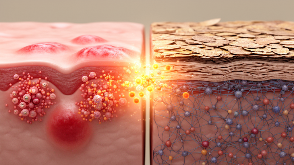
Chronic spontaneous urticaria (CSU) and atopic dermatitis (AD) are two of the most common chronic inflammatory skin diseases, and both are defined by severe, quality-of-life-destroying itch. Both draw on overlapping mast-cell-related biology, including IgE/FcεRI signaling, type 2 inflammation, neuroimmune signaling, and lipid mediator pathways. Yet clinically they usually look nothing alike: CSU produces transient wheals, flare reactions, and angioedema, while AD produces persistent eczematous lesions with chronic inflammation, excoriation, and lichenification. A new Perspective published in Frontiers in Allergy proposes a striking explanation for this discordance: the two diseases may represent different tissue interpretations of the same inflammatory vocabulary, with phenotypic divergence reflecting the tissue-response network in which mast cell activation is embedded rather than mediator identity alone.
The authors, Young-Min Ye of Ajou University School of Medicine in the Republic of Korea and Michihiro Hide of Hiroshima City Hiroshima Citizens Hospital and Hiroshima University in Japan, develop a mast-cell-centered comparative model rather than a comprehensive review of either disease. Epidemiologic data support a robust, at least partly bidirectional association between AD and CSU. Reported AD prevalence among CSU patients varies with study design and population, ranging broadly from approximately 10% to 40% and consistently exceeding rates in matched controls or the general population. Several cohorts report the risk of AD to be approximately 2- to 5-fold higher among CSU patients than matched controls, and a history of AD has been linked to increased subsequent risk of CSU, particularly when AD onset precedes CSU by several years. Comorbidity and temporal sequence of onset are established; temporal dissociation of disease activity within the same patient is not, because longitudinal studies concurrently measuring validated CSU and AD activity indices in the same patients are lacking.
The authors treat their clinical impression that severe eczema and severe wheals do not commonly peak together in the same patient as a hypothesis to be formally tested. If mediator identity alone determined phenotype, high mediator burden might be expected to produce phenotypic convergence. Instead, they propose that CSU and AD are best understood as distinct tissue-response programs. In CSU, mast-cell activation is wired to the dermal vascular/perivascular unit, producing edema, wheal, flare, and angioedema. In AD, mast-cell activation is embedded in a barrier-disrupted epithelial-alarmin-neuro-immune niche, producing chronic itch, scratching, keratinocyte activation, and eczematous inflammation. The model distinguishes feedback-limited vascular whealing from continuous neuro-epidermal inflammation.
In CSU, degranulation-ready mast cells and basophils are positioned within or near the microvascular unit, where FcεRI aggregation, IgG autoantibodies against IgE or FcεRI, IgE autoantibodies to self-antigens, MRGPRX2 ligands, coagulation/fibrinolysis-C5a signals, neuropeptides, and lipid mediators can converge. The preferential tissue-level output is transient vascular escape characterized by local histamine release, endothelial hyperpermeability with resultant plasma extravasation, wheal formation, angioedema, and rapid resolution. Of note, the threshold of histamine to induce visible flare (0.3–1 µM) is much less than that to induce wheal formation (3–10 µM), suggesting that wheal formation depends on a distinct, higher-threshold mechanism specific to urticaria rather than the lower-threshold neurogenic reflex underlying flare.
Mathematical modeling of CSU supports this vascular view. This work divides the wheal lifecycle into onset, development, and disappearance phases. Onset is driven by basophil-related endovascular positive feedback including the production of C5a from plasma complement C5, triggered by small stimuli on basophils and/or mast cells such as IgG autoantibodies against IgE/FcεRI, autoantigens binding to IgE, or tissue factor-expressing monocytes, and depends on the increase of exogenous coagulation potential. Development is related to mast cell-mediated vascular gap formation. Wheal disappearance is governed by an autoinhibitory purinergic mechanism: adenosine, generated through ATP breakdown in association with histamine release, suppresses further histamine release and tissue factor expression, terminating the positive-feedback loop that drives wheal expansion. Consistent with this self-limiting dynamic, histamine release appears in a short burst, further diluted by the plasma extravasation it helps generate. Together, these dynamics explain the rapid resolution characteristic of wheals.
Neuropeptides and lipid mediators may extend this vascular circuit beyond mast-cell histamine alone. Substance P (SP) can activate skin mast cells through MRGPRX2 and has also been linked to NK1R-mediated vasodilation and plasma extravasation, coupling sensory nerve input to the dermal mast cell-vascular unit. Both C5a and SP induce mast cell degranulation in the skin, but mast cells of other tissues show little or no responsiveness to these stimuli. Moreover, SP induces rapid release of TNF-α and IL-8 from human skin mast cells, but no significant levels of IL-4, IL-5, or IL-13. The release of mediators from human basophils induced by C5a largely depends on the environment, especially IL-3: C5a alone induces rapid release of histamine but little or no production of IL-4 by human basophils, despite promoting vigorous histamine release. However, basophils pretreated with IL-3 show large and long-lasting release of IL-4, IL-13, and LTC4 in response to C5a. Thus, in the absence of IL-3, activation of basophils and mast cells by C5a, SP, or transient IgE/FcεRI engagement mainly releases histamine with little type 2 cytokine production; in the presence of IL-3, the same stimuli can additionally drive type 2 cytokine release, favoring progression toward type 2 skin inflammation.
The role of platelet-activating factor (PAF) in this circuit is less settled. Human skin microdialysis studies found that PAF induces wheal-and-flare reactions without a measurable increase in dermal histamine or PGD2 release, unlike codeine-induced mast-cell activation, leading to the proposal that PAF drives endothelial hyperpermeability independently of mast-cell degranulation. However, in vitro studies show that PAF can directly induce degranulation of human mast cells, including LAD2 cells and induced pluripotent stem cell-derived mast cells. The authors suggest these findings need not conflict: if PAF triggers both mast-cell degranulation together with rapid endothelial leakage, plasma extravasation could dilute or disperse locally released histamine below the detection threshold of microdialysis sampling, consistent with burst-and-dilution dynamics. This proposed reconciliation is a hypothesis that has not been directly tested and would require simultaneous measurement of histamine, degranulation markers, and vascular permeability following PAF challenge. Together, these mechanisms explain why CSU lesions arise abruptly and resolve without persistent epidermal remodeling: the wheal, in this framework, is a feedback-regulated vascular event rather than a continuously expanding inflammatory plaque.
In AD, by contrast, mast cells are embedded in a prominent epidermal barrier-alarmin-neuroimmune circuit. Barrier disruption, microbial and allergen proteases, scratching injury, keratinocyte-derived TSLP, IL-33, and IL-25 combine with type 2 cytokines, sensory nerve activation, and mast-cell/immune-cell crosstalk to create a self-sustaining inflammatory niche. This multicellular network involves keratinocytes, diverse T-cell programs, group 2 innate lymphoid cells, dendritic cells, eosinophils, and other myeloid cells, with relative contributions varying across disease stages and endotypes. Within this broader network, mast cells may be preferentially conditioned toward inflammatory and neuroimmune amplification, although this does not exclude classical degranulation, protease release, or vascular effects. Human AD skin biopsy studies have demonstrated lesion-associated variation in mast cell numbers and tryptase/chymase phenotypes, increased mast cell-nerve contacts, altered chymase activity accompanied by increased mast cell IL-6 expression, and ultrastructural evidence of active secretion. A recent transcriptomic study further indicates that cytokine-conditioned human mast cells acquire distinct but overlapping functional programs; projection of these signatures onto AD skin single-cell data suggests heterogeneous IL-4/IL-13-, IFN-γ-, TGF-β-, and IL-33-associated mast cell states. These findings support context-dependent mast cell plasticity but do not establish a uniform AD-specific mast cell phenotype or demonstrate that any single mode of secretion predominates.
Rather than being expressed predominantly as an immediate vascular response, mast cell outputs in AD, including tryptase, cytokines, and chemokines, may be preferentially integrated into PAR2-related keratinocyte activation, neuronal sensitization, and neuroimmune itch amplification. Severe or chronic AD should not be reduced to a simple shift from type 2 to type 1 inflammation; a more appropriate model is immune broadening on a persistent type 2/Th22 background, with variable contributions from Th1, Th17, IL-1β, TNF, myeloid, microbial, and scratching-related signals. Within this multicellular program, mast cell outputs may contribute to chronic eczematous inflammation, excoriation, lichenification, and remodeling. Accordingly, inflammation-primed denotes a preferential functional tendency imposed by the AD tissue context, not a fixed mast cell subtype or an absence of degranulation. Neuropeptides illustrate shared biology with divergent tissue output: in both CSU and AD, sensory nerves release SP, calcitonin gene-related peptide (CGRP), and vasoactive intestinal peptide (VIP), which contribute to itch, neurogenic inflammation, mast-cell activation, vasodilation, and immune-cell recruitment. Serum SP is increased in CSU and correlates with disease activity, whereas in AD reported associations between circulating neuropeptides and disease severity have been less consistent, a pattern that may reflect a more chronic, locally regulated neuroepidermal circuit rather than an acute, circulating biomarker of disease activity. MRGPRX2 expression has also been reported in AD skin, and experimental evidence suggests that MRGPRX2-mediated tryptase release can amplify type 2 cytokine production, indicating that this pathway may also be incorporated into the chronic inflammatory network of AD, although its cellular distribution, endotype specificity, and relative contribution to human disease remain incompletely defined.
The same principle applies to coagulation, complement, protease/PAR signaling, and lipid mediators. In CSU, coagulation factor activation, thrombin and plasmin generation, fibrin turnover, and C5a/C5aR signaling are anatomically linked to the vascular/perivascular compartment, facilitating mast cell and basophil activation and endothelial leakage in the setting of increased coagulation potential, although the precise enzymatic mechanisms linking the coagulation, fibrinolysis, and complement cascades remain debated. In AD, protease-related and potentially coagulation-related signals may instead operate within a barrier-disrupted epidermal compartment through PAR2-expressing keratinocytes and sensory nerves, where their effects may be preferentially expressed as itch, epithelial inflammation, and remodeling rather than predominantly as vascular leakage. In AD, elevated circulating PAF has been reported, but its cellular routing and tissue-level consequences remain poorly defined. The network framework may also accommodate temporal plasticity between the two diseases, an extension the authors acknowledge remains speculative. One possibility is that a history of AD may leave a residual type 2/mast cell-primed milieu after overt eczema resolves, sustained by persistently elevated IgE, autoreactive IgE repertoires, eosinophil-biased inflammation, altered lipid mediator tone, neuroimmune sensitization, and residual barrier-immune memory; a second insult, such as autoimmunity, infection, stress, hormonal change, increase of coagulation potential, or IgE repertoire shift, may later shift this primed state toward dermal vascular whealing. The converse may also occur: in some patients with long-standing or treatment-refractory CSU, wheals are incompletely suppressed by antihistamines or omalizumab, allowing residual tissue inflammation to become more visible, with contributing signals including IL-13, IL-1β/IL-17A, eosinophilic or myeloid activation, PAF-related signaling, and scratching, consistent with the IL-13-dominant endotype previously identified in patients with delayed or absent response to omalizumab. Some patients may therefore acquire AD-like or eczematous inflammatory features; this does not mean that CSU becomes AD, but that the dominant tissue-response program may shift from vascular leakage toward tissue-retained inflammatory itch.
Several predictions are testable. Longitudinal registries should characterize the bidirectional temporal sequences of AD and CSU and serially assess validated disease-activity indices in patients with both conditions, using strict operational definitions, remission windows, diagnostic validation, and control for surveillance bias. Spatial profiling of CSU wheals and AD lesions should map mast cells, basophils, eosinophils, sensory nerves, endothelial activation, PAFR, TF/FVIIa-related coagulation signals, C5a/C5aR, PAR2, keratinocyte alarmins, and histamine or histamine-metabolizing pathways. Functional assays should test patient serum-primed mast cells, basophils, endothelial cells, and keratinocytes in parallel, determining whether CSU sera preferentially induce endothelial permeability and mast-cell degranulation whereas AD sera preferentially induce keratinocyte alarmins, cytokines, and neuroepidermal itch mediators. Finally, kinetic approaches to histamine, cytokines, and PAF biology are needed, because transient bursts, local dilution, and mediator dispersion may be missed by static measurements. The authors conclude that CSU and AD are not biologically incompatible diseases: they may represent different tissue interpretations of overlapping IgE-mast cell, neuropeptide, lipid mediator, protease, complement, and type 2 inflammatory signals. CSU is organized around a feedback-limited dermal neuropeptide-mast cell-vascular program that produces transient wheals and angioedema, while AD is organized around a disrupted barrier and a multicellular neuro-epidermal cytokine network that favors chronic eczematous inflammation. This tissue address may not be fixed: priming from prior AD and incomplete control of CSU both suggest that the dominant program can shift over time. Distinguishing shared mediators from their tissue address offers a testable framework for the clinical impression that wheals and eczema rarely reach peak severity together, and may guide biomarker discovery, endotype classification, and mechanism-based treatment selection, pending direct longitudinal and mechanistic validation.
Subject of Research: Shared mediators, divergent networks: a tissue-response hypothesis for discordant phenotypes in chronic spontaneous urticaria and atopic dermatitis
Article Title: Shared mediators, divergent networks: a tissue-response hypothesis for discordant phenotypes in chronic spontaneous urticaria and atopic dermatitis
Article References: Ye, Y.-M., & Hide, M. (2026). Shared mediators, divergent networks: a tissue-response hypothesis for discordant phenotypes in chronic spontaneous urticaria and atopic dermatitis. Frontiers in Allergy, 7, Article 1976519. https://doi.org/10.3389/falgy.2026.1976519
Image Credits: AI Generated
DOI: 10.3389/falgy.2026.1976519
Keywords: chronic spontaneous urticaria, atopic dermatitis, mast cells, MRGPRX2, neuro-immune inflammation, vascular permeability, platelet-activating factor, complement-coagulation crosstalk, Shared, mediators, divergent, networks
Cite Scienmag News
Dean Parker. (October 15, 2026). Why hives and eczema rarely flare together: shared mediators, different tissue networks. Scienmag. https://scienmag.com/why-hives-and-eczema-rarely-flare-together-shared-mediators-different-tissue-networks/
Dean Parker. "Why hives and eczema rarely flare together: shared mediators, different tissue networks." Scienmag, 15 October 2026, https://scienmag.com/why-hives-and-eczema-rarely-flare-together-shared-mediators-different-tissue-networks/. Accessed 15 October 2026.
Dean Parker. "Why hives and eczema rarely flare together: shared mediators, different tissue networks." Scienmag. October 15, 2026. https://scienmag.com/why-hives-and-eczema-rarely-flare-together-shared-mediators-different-tissue-networks/


