A 51-year-old woman in China lost her newly implanted aortic valve prosthesis to a catastrophic migration five months after transcatheter aortic valve replacement, and that failure — occurring without infection or any identifiable procedural error — is what finally led doctors to the diagnosis that had eluded them for years: Behçet disease-associated aortitis. The case, published as a case report in Frontiers in Cardiovascular Medicine on 28 October 2026, is described by its authors as the first reported instance of late TAVR prosthesis migration attributed to Behçet-related aortitis.
The story began two years before her admission to West China Hospital in Chengdu, when the woman developed multifocal bone pain and eventually became unable to walk because of severe lower back pain. Imaging at an outside facility had shown multiple osteolytic lesions, and a clavicular biopsy revealed bone marrow edema reported only as suggestive of chronic osteomyelitis. She also tested positive on a T-SPOT.TB assay and a strongly positive purified protein derivative skin test, so she received empiric anti-tuberculosis therapy. Her symptoms worsened after a month of that treatment, prompting transfer to the Chinese hospital.
At evaluation she reported shortness of breath on mild exertion, classified as New York Heart Association functional class II–III, along with recurrent oral ulcers. Examination found multiple acneiform lesions on her face, a diastolic murmur at the aortic valve area, percussion tenderness over the lumbar spine and ribs, and mild bilateral lower extremity edema. She had an eight-year history of essential hypertension. Laboratory testing showed microcytic anemia with a hemoglobin of 80 g/L and a profound inflammatory response: an erythrocyte sedimentation rate above 120 mm/h, C-reactive protein of 77 mg/L, serum amyloid A of 149 mg/L, and alpha-1 acid glycoprotein of 1,920 mg/L. Testing for multiple myeloma, hyperparathyroidism, major autoimmune rheumatic diseases and autoimmune hepatitis came back negative, as did HLA-B27, HLA-B51 and the pathergy test.
Computed tomography revealed hyperostosis and sclerosis of the right sternoclavicular and first sternocostal joints, together with multifocal spinal involvement from the cervical through the lumbar spine, including facet joint erosion and partial vertebral fusion. Whole-body bone scintigraphy confirmed multifocal radiotracer uptake in the sternum, the right sternoclavicular joint and multiple vertebrae. Ankylosing spondylitis was excluded by negative HLA-B27 typing and the absence of sacroiliitis or syndesmophytes. The combination of anterior chest wall involvement, multifocal osteolytic lesions and acne fulfilled the diagnostic criteria proposed by Benhamou and colleagues, and the team made a clinical diagnosis of SAPHO syndrome — synovitis, acne, pustulosis, hyperostosis and osteitis. Her International Criteria for Behçet Disease score at that stage was only 3 points, from recurrent oral aphthosis and acneiform lesions, below the 4-point threshold, so Behçet disease was considered unlikely.
Transthoracic echocardiography, prompted by her heart failure symptoms, revealed severe aortic regurgitation with a vena contracta of 7 mm, a dilated left ventricle with an end-diastolic dimension of 60 mm, and preserved systolic function with an ejection fraction of 58 percent. After a month of diclofenac, which reduced her pain score from 7 to 2, she was readmitted with worsening heart failure. The team chose transcatheter aortic valve replacement over open surgery specifically because sternotomy, they feared, might impair bone healing in the setting of suspected SAPHO-related active osteitis. Pre-procedure CT angiography showed a tricuspid aortic valve with a 23.8 mm annulus, a 31.3 mm average sinus diameter, a 27.3 mm left ventricular outflow tract, and no root calcification.
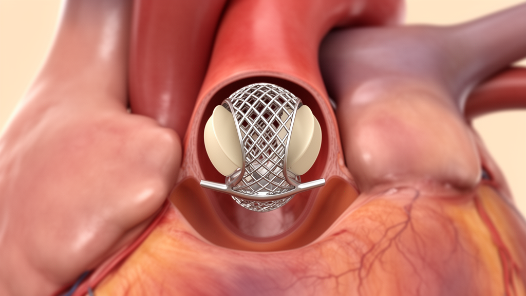
The transapical procedure used the J-valve system, a self-expanding prosthesis designed for aortic regurgitation without annular calcification. The device fixes itself with three U-shaped nitinol graspers deployed within the aortic sinuses, which also protect the coronary ostia from obstruction. A 25-mm valve was deployed and seated within the graspers, and intraoperative transesophageal echocardiography and aortic root angiography confirmed optimal position, normal leaflet function, no paravalvular leak and stable anchorage. She was discharged five days later on warfarin.
Three months after the procedure she returned with dizziness and limb weakness. Cerebral MRI showed a small acute ischemic lesion in the right occipital lobe, and CT angiography identified a thrombus in the superior mesenteric artery — arterial thromboses that developed despite anticoagulation, with an international normalized ratio of 1.9. Echocardiography at that visit showed normal prosthetic valve function with no paravalvular leak, and CT confirmed stable valve positioning. She also had second-degree type II atrioventricular block, for which a dual-chamber pacemaker was implanted. Two months later, at five months post-TAVR, she presented with chest tightness and fatigue. Imaging showed the prosthesis had migrated about 10 mm below the native annular plane into the left ventricular outflow tract, producing severe paravalvular regurgitation, while the prosthesis itself remained structurally intact with normal leaflet function. CT also showed aortic wall thickening consistent with aortitis. There was no evidence of infective endocarditis: blood cultures were repeatedly negative and no vegetations were seen.
That constellation — recurrent oral ulcers, acneiform lesions, osteolytic bone lesions, arterial thrombosis and prosthetic valve dehiscence without infection — led the team to a revised diagnosis of Behçet disease-associated aortitis. The cerebral infarction and mesenteric artery thrombosis added a point for vascular involvement, bringing her ICBD score to 4 and meeting the diagnostic threshold, though the authors note these features were not synthesized into a unifying diagnosis until the migration occurred. To reduce the risk of suture dehiscence from active vasculitis, she first received a three-month course of prednisone, thalidomide and cyclophosphamide, which significantly reduced inflammatory markers. She then underwent a Bentall procedure with a 23-mm mechanical valved conduit. Intraoperatively the aortic annulus appeared friable and inflamed, and histopathology of the excised aortic root tissue showed uneven valvular thickening, focal myxoid degeneration, lymphocytic infiltration and focal necrosis on the intimal surface, without infective organisms, granulomatous inflammation, storiform fibrosis or IgG4-positive plasma cells — findings the authors say supported a sterile inflammatory etiology while arguing against infective endocarditis and IgG4-related disease, though they acknowledge such findings are not specific for Behçet aortitis.
The discussion places the case within a broader and sobering surgical literature. The authors cite a report by Han and colleagues in which only 2 of 15 patients with surgically confirmed Behçet-related aortic regurgitation had been diagnosed with Behçet disease before their first cardiac operation; the remaining 13 were identified only after characteristic postoperative complications, most commonly prosthetic valve dehiscence. In the largest surgical series to date, by Choi and colleagues, only 17.1 percent of 35 patients met International Study Group criteria preoperatively, roughly 25 percent remained undiagnosed by either ISG or ICBD criteria even at surgery, and the two-year incidence of paravalvular leak in Behçet patients was 51.4 percent compared with approximately 1 percent in non-Behçet controls. Patients with incomplete-criteria Behçet disease fared paradoxically worse precisely because the condition went unrecognized and postoperative immunosuppression was delayed. The authors also propose a mechanism specific to transcatheter valves: unlike balloon-expandable valves anchored by radial force against a calcified annulus, the J-valve depends critically on the integrity of the aortic sinus wall, and inflammation-mediated compromise of that tissue may have displaced the entire stent-valve complex.
Over the following two years the woman remained asymptomatic with no recurrence of bone pain and no tuberculous reactivation despite substantial prior immunosuppressive exposure. Serial echocardiography showed sustained reverse remodeling, with the ejection fraction improving from 58 to 67 percent and the left ventricular end-diastolic dimension decreasing from 60 to 48 mm, and the mechanical valve functioned normally with a peak forward flow velocity of 2.3 m/s and a mean gradient of 11 mmHg at the latest follow-up. The authors acknowledge that current guidelines favor surgical aortic valve replacement for a 51-year-old patient and that their initial rationale rested on an incomplete diagnostic framework; in retrospect, they write, the optimal pathway would have been early recognition of a unifying inflammatory etiology, induction of remission through immunosuppression, and definitive aortic root replacement. Their conclusion is directed at clinicians: when unexplained aortic regurgitation coexists with systemic inflammatory features, a unifying diagnosis should be pursued and disease control established before structural cardiac intervention, and prosthetic valve dehiscence in the absence of infection should raise strong suspicion for inflammatory aortitis even when formal classification criteria are initially unmet. As a single case report, the findings cannot establish how often such migration occurs or how reliably it signals Behçet disease, but they add a striking example to the documented pattern of delayed cardiovascular diagnosis in this rare vasculitis.
Subject of Research: Prosthetic valve migration after transcatheter aortic valve replacement unmasking Behçet disease-associated aortitis in a single case report
Article Title: Case Report: Prosthetic valve migration unmasking behçet disease after transcatheter aortic valve replacement
Article References: Wang, X., Qin, Y., & Bai, W. (2026). Case Report: Prosthetic valve migration unmasking behçet disease after transcatheter aortic valve replacement. Frontiers in Cardiovascular Medicine, 13, 1947155. https://doi.org/10.3389/fcvm.2026.1947155
Image Credits: AI Generated
DOI: 10.3389/fcvm.2026.1947155
Keywords: Behçet disease, aortitis, TAVR, prosthesis migration, aortic regurgitation, SAPHO syndrome, Bentall procedure, paravalvular leak, vasculitis, case report, J-valve, cardiovascular medicine
Cite Scienmag News
Ophelia Keating. (October 28, 2026). Valve Migration After TAVR Unmasked Hidden Behçet Disease in One Patient. Scienmag. https://scienmag.com/valve-migration-after-tavr-unmasked-hidden-behcet-disease-in-one-patient/
Ophelia Keating. "Valve Migration After TAVR Unmasked Hidden Behçet Disease in One Patient." Scienmag, 28 October 2026, https://scienmag.com/valve-migration-after-tavr-unmasked-hidden-behcet-disease-in-one-patient/. Accessed 28 October 2026.
Ophelia Keating. "Valve Migration After TAVR Unmasked Hidden Behçet Disease in One Patient." Scienmag. October 28, 2026. https://scienmag.com/valve-migration-after-tavr-unmasked-hidden-behcet-disease-in-one-patient/