


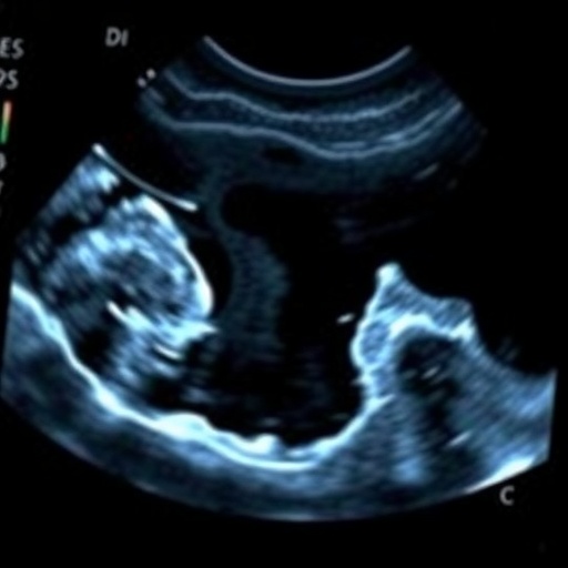
In a groundbreaking study published in the Journal of Perinatology on May 27, 2026, researchers have unveiled a novel bedside ultrasound imaging technique designed to measure muscle cross-sectional area in both preterm and term infants. This pioneering approach harnesses the power of non-invasive imaging to provide critical insights into neonatal muscle health without the customary reliance on more invasive or less feasible methods in delicate patient populations. The implications for neonatal care, growth monitoring, and nutritional assessment could mark a paradigm shift in how clinicians address early-life muscular development.
The study, led by Yeager, S., Hendrixson, D.T., Cunha, G.M., and colleagues, specifically targeted the feasibility of applying bedside ultrasound methods within neonatal intensive care units (NICUs). Unlike traditional imaging methodologies that often require transportation of fragile infants or exposure to ionizing radiation, bedside ultrasound is portable, safe, and repeatable. Its adoption promises to overcome several logistical and ethical challenges inherent in neonatal muscle assessment while enabling real-time data acquisition that can inform clinical decision-making instantaneously.
At the core of this investigation lies the measurement of muscle cross-sectional area (CSA), a biomarker directly correlated with muscle mass and indirectly with overall growth and nutritional status in infants. Muscle CSA has been historically difficult to assess accurately in neonates due to their small size, sensitivity, and the technical limitations of standard imaging tools. By adapting ultrasonographic protocols to the neonatal context, the research team has created a reliable metric that can be obtained without disrupting critical care routines.
Muscle mass in infancy is a crucial determinant of long-term health outcomes. Preterm infants, in particular, face numerous challenges related to inadequate protein accumulation and developmental delays, which can translate into compromised motor function and metabolic disorders later in life. This study addresses a significant clinical gap: the absence of simple, accurate means to monitor muscle growth in this vulnerable cohort. By facilitating bedside assessments, healthcare providers can tailor nutritional interventions more precisely, potentially mitigating long-term adverse effects.
The research design involved a cohort of both preterm and term infants, thereby allowing for comparative analyses that underscore the method’s versatility. Ultrasound imaging was performed using high-frequency linear transducers suitable for small anatomical structures. This technical adaptation enabled the acquisition of high-resolution images from specific muscle groups, such as the quadriceps and biceps brachii, to quantify their CSA accurately. The feasibility study focused on operational protocols, including optimal probe placement, imaging parameters, and operator training requirements.
An important technical consideration addressed by the authors is image reproducibility and measurement reliability. Neonatal ultrasound poses unique challenges such as movement artifacts, limited acoustic windows, and the necessity for non-sedated procedures. The investigators employed rigorous intra- and inter-rater reliability testing, deploying standardized measurement techniques and calibration protocols. The results demonstrated high consistency across operators and sessions, bolstering confidence in the clinical utility of the ultrasound-based measurement approach.
This study also delved into the biophysical principles underpinning ultrasound imaging in neonates. The reflective properties of muscle tissue, combined with the relative homogeneity of fatty infiltration in infants, influence the echogenicity and contrast critical for delineating anatomical boundaries. By optimizing gain settings and frequency ranges, the researchers enhanced delineation of muscle borders, ensuring accurate CSA calculations. Such technical refinements are essential for translating experimental imaging into routine clinical practice.
The implications of this technique extend beyond mere measurement; they open avenues for dynamic monitoring of muscle growth trajectories. Repeated ultrasound assessments can track changes in muscle CSA longitudinally, providing a sensitive surrogate for nutritional adequacy, metabolic health, and response to interventions such as physiotherapy or pharmacotherapy. This real-time monitoring capability represents a significant advance over static growth charts or indirect biomarkers reliant on systemic parameters.
From a clinical workflow perspective, the portability and speed of bedside ultrasound facilitate its integration into NICU routines without imposing additional stress or risk on neonates. The non-invasive nature aligns with principles of minimizing harm and promoting comfort in fragile infants. Moreover, the digital imaging outputs enable seamless documentation, remote consultation, and longitudinal data archiving, fostering multidisciplinary collaboration and evidence-based care pathways.
The study’s innovative nature was further highlighted by its potential to inform research on neonatal muscle biology. By providing a practical tool to quantify muscle morphology in vivo, researchers can investigate correlations with genetic markers, environmental influences, and developmental trajectories. This opens prospects for uncovering mechanisms underlying neonatal diseases linked to muscle deficits, such as neuromuscular disorders or intrauterine growth restriction.
Technologically, the advancement taps into ongoing improvements in ultrasound transducer miniaturization and image processing software. Real-time edge detection algorithms, automated delineation, and 3D reconstruction techniques could further enhance the precision and clinical applicability of muscle CSA measurement. The study serves as a foundational step encouraging the development of integrated devices tailored specifically for neonatal bedside muscular assessment.
The multidisciplinary composition of the research team, involving neonatologists, radiologists, biomedical engineers, and physiologists, reflects the complexity and translational importance of the work. Their collaborative efforts ensured that the technique is not only scientifically valid but also clinically feasible and replicable across various care settings. Such integrative approaches are vital for bridging the gap between innovative diagnostics and improved patient outcomes.
While this feasibility study lays the groundwork, the authors acknowledge the need for expansive longitudinal studies to validate muscle CSA measurements against clinical endpoints such as functional development and long-term health. Further exploration of normative data stratified by gestational age, sex, and ethnicity will be necessary to refine interpretation criteria. Additionally, incorporation into multicenter trials would establish robustness and facilitate widespread adoption.
Beyond individual patient care, this technique holds promise for monitoring populations at risk of malnutrition or muscle wasting in resource-limited settings. The affordability and portability of ultrasound devices, when combined with standardized protocols, offer a practical alternative to expensive imaging modalities. This democratization of muscle assessment could impact global health initiatives aimed at reducing neonatal morbidity and mortality.
Fundamentally, this research reimagines neonatal care through the lens of precision phenotyping. Muscle mass assessment via bedside ultrasound embodies a shift from reactive treatment to proactive surveillance, empowering caregivers with timely, actionable data. As neonatal medicine advances toward personalized interventions, such innovations will be instrumental in shaping outcomes from the earliest stages of life.
In summary, the unveiling of bedside ultrasound imaging for quantifying muscle cross-sectional area in preterm and term infants represents a critical milestone in neonatal diagnostics. This technique harmonizes technological innovation with clinical pragmatism, fostering improved assessment of muscle health with direct translational applications. The continued evolution and validation of this approach will undoubtedly influence neonatal care standards and open new horizons for research and therapeutic strategies worldwide.
Cite Scienmag News
Harold Sullivan. (May 27, 2026). Ultrasound Measures Muscle Size in Preterm Infants. Scienmag. https://scienmag.com/ultrasound-measures-muscle-size-in-preterm-infants/
Harold Sullivan. "Ultrasound Measures Muscle Size in Preterm Infants." Scienmag, 27 May 2026, https://scienmag.com/ultrasound-measures-muscle-size-in-preterm-infants/. Accessed 4 September 2026.
Harold Sullivan. "Ultrasound Measures Muscle Size in Preterm Infants." Scienmag. May 27, 2026. https://scienmag.com/ultrasound-measures-muscle-size-in-preterm-infants/


