


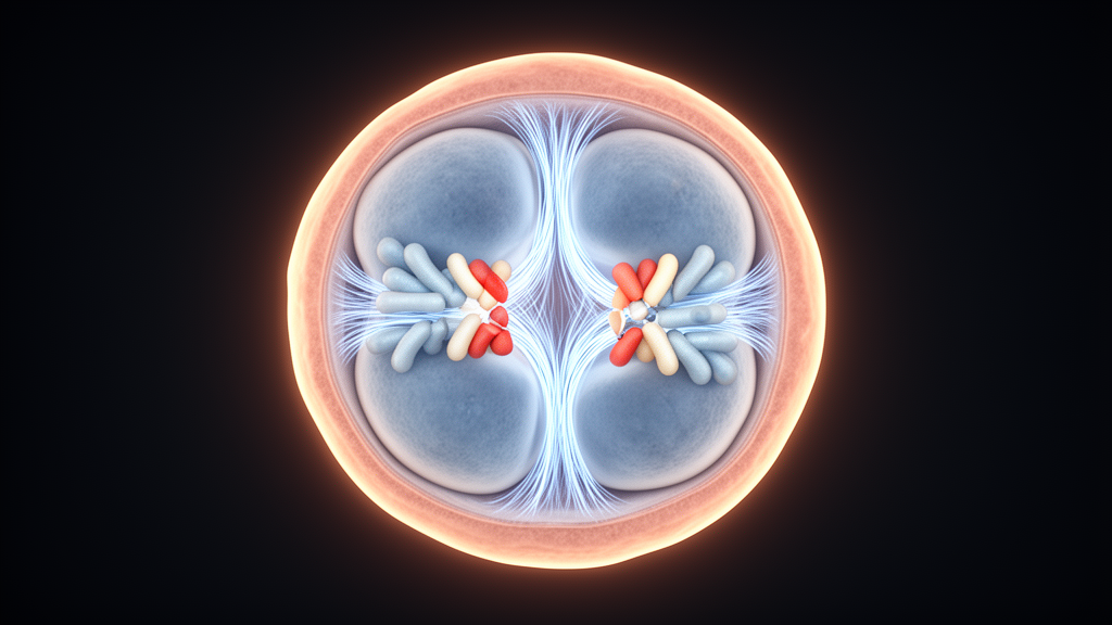
Carriers of balanced autosomal reciprocal translocations usually feel perfectly healthy, yet their reproductive lives can be dominated by miscarriage, infertility, or the birth of children with serious congenital malformations. The reason lies in meiosis. When two non-homologous chromosomes have swapped fragments, the derivative chromosomes and their normal homologues gather into a four-chromosome structure called a quadrivalent during the first meiotic division. How that quadrivalent segregates determines whether the resulting gamete is genetically balanced or carries a large, often lethal, chromosomal imbalance. A new retrospective study published in Comparative Cytogenetics has now dissected which features of the quadrivalent steer this process in human embryos, and how natural selection filters the outcomes by the blastocyst stage.
The research team, working at the International Center for Reproductive Medicine, analyzed 39 couples in which one partner carried an autosomal reciprocal translocation and who underwent in vitro fertilization with preimplantation genetic testing for structural rearrangements between 2016 and 2021. The dataset comprised 306 cleavage-stage embryos, examined on day three, and trophectoderm cells from 93 blastocyst-stage embryos, examined on day five or six. Genetic testing relied on fluorescent in situ hybridization, next-generation sequencing, or array comparative genomic hybridization, allowing the researchers to infer which segregation mode had produced each embryo. Karyotypes were confirmed on peripheral blood lymphocytes, and all participants signed informed consent.
Across all 399 embryos, alternate segregation, the only mode that yields genetically normal or balanced gametes, was the most common pattern, detected in 32 percent of cases. Adjacent-1 segregation followed at 24 percent, the 3:1 mode at 21 percent, adjacent-2 at 13 percent, and the rare 4:0 pattern in just 1 percent. In 9 percent of embryos the segregation mode could not be determined because of mosaicism or polyploidy, and these were excluded from further analysis. The overall picture confirms that even though alternate segregation dominates, carriers still produce a strikingly high proportion of unbalanced gametes, which underlies their elevated risk of reproductive failure.
The most dramatic finding emerged when the researchers compared day-three and day-five/six embryos. The frequency of 3:1 segregation collapsed from 28 percent at the cleavage stage to just 4 percent at the blastocyst stage, a difference that was highly statistically significant with a p-value below 0.0001. Alternate segregation rose from 34 to 45 percent and adjacent-1 from 24 to 33 percent, though these increases did not reach statistical significance, likely because of the modest sample size. The explanation, the authors argue, is natural selection: embryos produced by 3:1 segregation carry tertiary or interchange trisomies and monosomies, which represent far larger chromosomal imbalances than the partial trisomies and monosomies of adjacent-1 products, and such heavily unbalanced embryos rarely survive to the blastocyst stage.
To understand what drives pathological segregation in the first place, the team turned to cleavage-stage embryos, where the embryonic genome has not yet triggered substantial selection, and classified each translocation by three quadrivalent features. The degree of asymmetry was quantified by measuring translocated and centric segment lengths with the UCSC genome browser on the GRCh38/hg38 assembly, and calculating the ratio of the longest to the shortest segment of each type; ratios of two or more in both categories defined severe asymmetry. Terminal breakpoints were defined as breakpoints falling within the distal 20 percent of a chromosome arm. Acrocentric involvement was recorded whenever chromosome 13, 14, 15, 21, or 22 participated in the rearrangement.
Of the 39 translocations, 28 showed mild asymmetry and 11 severe asymmetry, but asymmetry degree had no detectable effect on the frequency of any segregation mode at the cleavage stage. Acrocentric involvement, present in 18 cases, shifted the balance between pathological modes: it increased the frequency of adjacent-2 segregation and reduced adjacent-1, without altering the production of balanced embryos. The decisive factor, however, was the presence of terminal breakpoints, found in seven translocations. Embryos from carriers with terminal breakpoints showed a significantly higher frequency of 3:1 segregation, with a p-value of 0.002, while the rate of balanced embryo formation remained unchanged.
The viability analysis revealed an unexpected twist. When embryos from carriers with terminal breakpoints were followed to the blastocyst stage, significantly more embryos derived from alternate segregation and, strikingly, from adjacent-1 segregation survived, with p-values of 0.0513 and 0.0007 respectively. The authors attribute this to the small size of the translocated segments in rearrangements with terminal breakpoints. Adjacent-1 products carry partial monosomy or trisomy of the translocated segments only, and when those segments are tiny, the resulting imbalance may fall within the limits compatible with implantation and development. Classical models by Daniel and later Cohen estimated that viable imbalances rarely exceed a few percent of the haploid autosome length, and most adjacent-1 embryos in this cohort fit within those bounds.
This survival effect carries a clinical warning. An embryo that reaches the blastocyst stage after adjacent-1 segregation may look developmentally competent, yet it can harbor an imbalance capable of producing a child with multiple congenital malformations. The findings therefore support preimplantation genetic testing of blastocyst-stage embryos as preferable to cleavage-stage testing for translocation carriers, since blastocyst testing both benefits from natural selection against severely unbalanced embryos and provides a broader genomic assessment. At the same time, the study shows that quadrivalent asymmetry and acrocentric involvement, which earlier work had implicated in segregation patterns, do not appear to influence embryo survival by the blastocyst stage regardless of the preceding segregation mode.
The authors acknowledge limitations, including the small sample size and the fact that only a limited number of chromosomes were analyzed in cleavage-stage embryos, meaning some selection by the blastocyst stage could reflect aneuploidy of chromosomes outside the rearrangement. Even so, the work delivers a coherent picture: terminal breakpoints predispose to 3:1 segregation during gametogenesis, natural selection removes most 3:1 products by day five or six, and the same terminal breakpoints paradoxically enhance the survival of adjacent-1 embryos whose small imbalances may still cause congenital disease. For the estimated one in five hundred people who carry a reciprocal translocation, these patterns promise sharper risk assessment and better-informed counseling in IVF programs.
Subject of Research: Chromosome segregation patterns in human preimplantation embryos from reciprocal translocation carriers
Article Title: Assessment of quadrivalent characteristics influencing chromosome segregation by analyzing human preimplantation embryos from reciprocal translocation carriers
Article References: Tonyan, Z. N., Puppo, I. L., Saifitdinova, A. F., Vavilova, T. V., & Glotov, A. S. (2024). Assessment of quadrivalent characteristics influencing chromosome segregation by analyzing human preimplantation embryos from reciprocal translocation carriers. Comparative Cytogenetics, 18, 1-13. https://doi.org/10.3897/compcytogen.18.115070
Image Credits: AI Generated
DOI: 10.3897/compcytogen.18.115070
Keywords: reciprocal translocation, quadrivalent, chromosome segregation, preimplantation genetic testing, blastocyst, meiosis, terminal breakpoints, acrocentric chromosomes, IVF, PGT-SR, Assessment, characteristics
Cite Scienmag News
Drew Townsend. (October 18, 2026). Terminal Breakpoints Shape Chromosome Segregation and Embryo Survival in Translocation Carriers. Scienmag. https://scienmag.com/terminal-breakpoints-shape-chromosome-segregation-and-embryo-survival-in-translocation-carriers/
Drew Townsend. "Terminal Breakpoints Shape Chromosome Segregation and Embryo Survival in Translocation Carriers." Scienmag, 18 October 2026, https://scienmag.com/terminal-breakpoints-shape-chromosome-segregation-and-embryo-survival-in-translocation-carriers/. Accessed 18 October 2026.
Drew Townsend. "Terminal Breakpoints Shape Chromosome Segregation and Embryo Survival in Translocation Carriers." Scienmag. October 18, 2026. https://scienmag.com/terminal-breakpoints-shape-chromosome-segregation-and-embryo-survival-in-translocation-carriers/


