

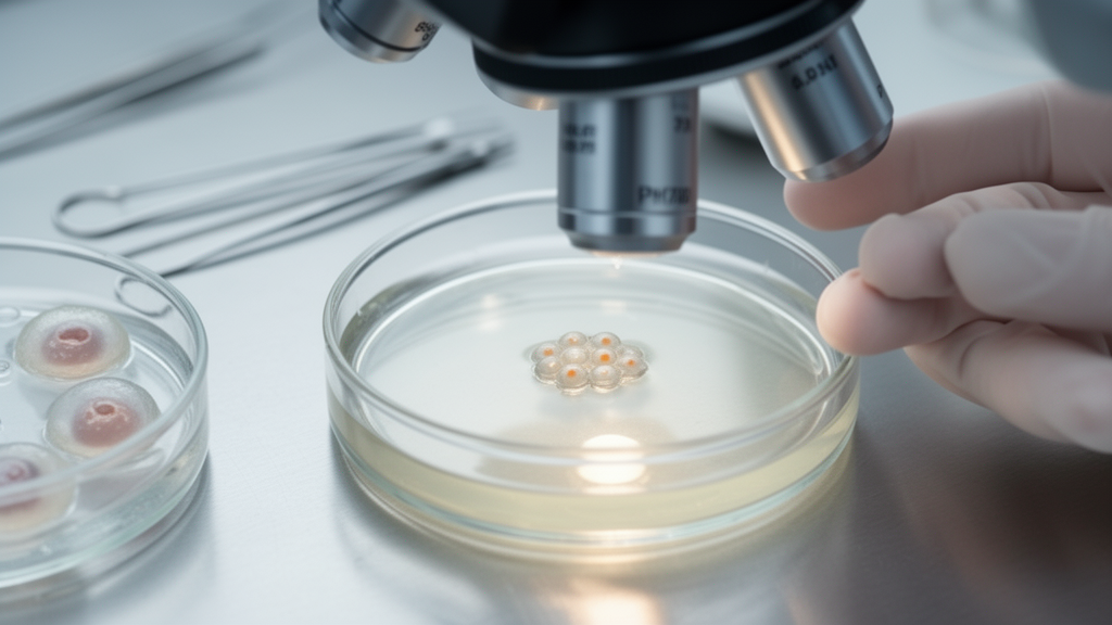
Embryos produced from ovarian follicles smaller than 8 millimetres reached the start and completion of blastulation about eight hours later than embryos derived from follicles of at least 11 millimetres in a small retrospective time-lapse analysis of intracytoplasmic sperm injection cycles. The delay was confined to the transition into blastocyst development: early pronuclear and cleavage timings did not differ significantly, nor did the interval between the onset of blastulation and full blastocyst formation. The study included 62 blastocysts from 18 women, including 20 from small follicles and 42 from larger follicles, and was not designed to demonstrate that other outcomes were equivalent. Although five of 10 tested blastocysts from small follicles were euploid, and two frozen-thawed transfers of euploid small-follicle embryos produced live births, the chromosomal and clinical samples were too small for reliable comparative estimates.
Researchers at Acibadem Maslak Hospital in Istanbul analysed embryos monitored continuously in an EmbryoScope+ incubator between March and August 2025. Follicle diameter was measured on the retrieval day as the mean of two perpendicular dimensions, with a three-millimetre gap separating the two analytical groups: follicles smaller than 8 millimetres and those measuring at least 11 millimetres. Follicles in the intermediate 8–11 millimetre range were excluded. All morphokinetic annotations were made by one experienced embryologist using established criteria, with time zero defined as the moment of sperm injection. Because several women contributed embryos to both size groups, the investigators used linear mixed models with a random intercept for patient and generalized estimating equations for binary outcomes, allowing repeated embryos from the same woman to remain clustered rather than being treated as independent observations.
The clearest timing difference appeared at blastulation. The median time to the start of blastulation was 101.2 hours after injection for embryos from follicles smaller than 8 millimetres, compared with 95.3 hours for those from follicles of at least 11 millimetres, a nominal difference of 5.9 hours in the unadjusted comparison. Full blastocyst formation occurred at a median of 108.1 hours versus 104.3 hours. Mixed-model estimates accounting for within-patient clustering placed the adjusted delay at 7.91 hours for blastulation onset and 8.29 hours for full blastocyst formation, with both confidence intervals excluding zero. Substantial patient-level variation was present: intraclass correlation coefficients of 0.343 and 0.308 indicated that roughly one-third of the variation in these timings was attributable to differences between women.
The result was not driven by the women who supplied embryos to only one group. Among seven patients who contributed blastocysts from both follicle-size categories, the estimated delay was 8.72 hours for the start of blastulation and 10.16 hours for full blastocyst formation. It also remained similar after statistical adjustment for maternal age, male-factor infertility and trigger protocol, and after excluding the three couples with male-factor infertility. A mediation analysis indicated that the group difference was established at the onset of blastulation rather than accumulating later: once timing of blastulation onset was included, the direct effect on full blastocyst formation was not significant. The interval between these two events had medians of 8.6 hours for small-follicle embryos and 7.9 hours for larger-follicle embryos, a difference that was not statistically significant.
Not every developmental measure favoured the larger-follicle group. Among all oocytes retrieved, 51.4% from follicles smaller than 8 millimetres were at metaphase II, compared with 74.3% from follicles of at least 11 millimetres, indicating that the small-follicle group was less mature at collection. Among oocytes that had reached metaphase II, however, the fertilization rate was higher in the small-follicle group: 92.9%, compared with 75.3% after intracytoplasmic sperm injection. The day-5 blastocyst rate per fertilized oocyte was lower from small follicles, at 28.8% versus 49.3%, while the day-6 rate was 9.6% versus 7.2%. The combined blastocyst formation rate was 38.5% versus 56.5%, but that difference was not significant after accounting for patient clustering. These figures were exploratory, and their wide confidence intervals mean that the study cannot establish that the groups were equivalent.
Culture system complicated interpretation of the overall blastocyst comparison. Every embryo from a follicle smaller than 8 millimetres was placed in the time-lapse incubator, whereas embryos from larger follicles were distributed between time-lapse and conventional incubation. Within the larger-follicle group, blastocyst formation per injected oocyte was lower in the time-lapse system than in conventional incubators, 32.6% versus 50.0%. When the comparison was restricted to embryos cultured in the time-lapse system, the blastocyst rate was 35.7% for small-follicle embryos and 32.6% for larger-follicle embryos, with no significant difference. Because allocation was not randomized and was associated with follicle group, the investigators could not attribute the full between-group blastocyst difference to follicle size alone. They considered the incubator disparity more likely a consequence of non-random allocation than an effect of time-lapse culture itself.
Preimplantation genetic testing was performed in seven patients who had a history of recurrent pregnancy loss. Of 10 biopsied blastocysts derived from follicles smaller than 8 millimetres, five were euploid, one was mosaic and four were aneuploid; seven of 23 blastocysts from larger follicles were euploid. The difference in euploidy rates, 50.0% versus 30.4%, was not statistically significant. This comparison was based on only 33 tested embryos and was heavily influenced by one patient, who contributed 14 of the 23 larger-follicle blastocysts. Excluding that patient produced similar rates in the remaining samples, 55.6% versus 44.4%. The study therefore provides no reliable evidence for a difference in euploidy, but its small, non-representative sample also cannot establish the absence of one.
The clinical observations were encouraging but highly selected. When suitable embryos derived from follicles of at least 11 millimetres were available, they were preferred for transfer. Frozen-thawed single-embryo transfers using euploid blastocysts from follicles smaller than 8 millimetres occurred in two patients who had no such alternative, and both resulted in live births. These two events demonstrate that small-follicle embryos can produce euploid blastocysts and can result in live birth, but they cannot estimate implantation or live-birth probabilities. The researchers emphasized that the transfer group was small and non-random, while the selection of larger-follicle embryos whenever possible prevents direct comparison of clinical outcomes between follicle-size categories.
The findings suggest that follicle origin may influence the timing of blastulation without establishing a corresponding loss of developmental potential. Embryos from very small follicles had completed early cleavage on a similar schedule, then entered blastulation later and proceeded from blastulation onset to full expansion at a comparable pace. One possible explanation is that oocytes from less-developed follicles have had less time to accumulate maternal RNA, proteins, mitochondria or metabolic support, making a difference more apparent when the energetic demands of blastocyst expansion increase. None of those cellular mechanisms was measured, however, so this remains a hypothesis rather than a demonstrated explanation. The retrospective design, single-centre setting, 18-patient sample, absence of an a priori power calculation, categorical rather than exact per-oocyte follicle measurements, unblinded annotation and partial confounding with culture system limit generalization. Larger prospective studies would be required before the results could guide clinical practice or embryo-selection algorithms.
Subject of Research: Association between ovarian follicle size and blastocyst morphokinetic timing in an ICSI cohort
Article Title: Embryos derived from small follicles (<8 mm) exhibit delayed blastulation: a retrospective time-lapse analysis
Article References: Peker, N., Yücetürk, A., Karaosmanoğlu, Ö., Elmas, B., Özer Aslan, İ., & Tıraş, B. (2026). Embryos derived from small follicles (<8 mm) exhibit delayed blastulation: a retrospective time-lapse analysis. Frontiers in Endocrinology, 17, Article 1980066. https://doi.org/10.3389/fendo.2026.1980066
Image Credits: AI Generated
DOI: 10.3389/fendo.2026.1980066
Keywords: small ovarian follicles, blastulation, blastocysts, time-lapse imaging, ICSI, morphokinetics, euploidy, assisted reproduction, ovarian reserve, Embryos, derived, small
Cite Scienmag News
Scienmag. (October 20, 2026). Small-follicle embryos show delayed blastulation in retrospective ICSI study. https://scienmag.com/small-follicle-embryos-show-delayed-blastulation-in-retrospective-icsi-study/
Scienmag. "Small-follicle embryos show delayed blastulation in retrospective ICSI study." Scienmag, 20 October 2026, https://scienmag.com/small-follicle-embryos-show-delayed-blastulation-in-retrospective-icsi-study/. Accessed 20 October 2026.
Scienmag. "Small-follicle embryos show delayed blastulation in retrospective ICSI study." Scienmag. October 20, 2026. https://scienmag.com/small-follicle-embryos-show-delayed-blastulation-in-retrospective-icsi-study/


