


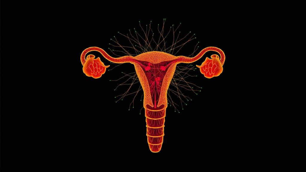
One of the most striking events in vertebrate embryology unfolds in near silence: the paired Müllerian ducts, which have the potential to become the female reproductive tract, either mature into oviducts or dissolve almost entirely, depending on the sex of the embryo. In mammals this process has been studied for decades, but the chicken embryo offers something mammals cannot — a system in which both ducts are accessible, sexually dimorphic fates play out on a predictable schedule, and, uniquely among birds commonly studied in the laboratory, the female embryo itself performs an asymmetric developmental maneuver, retaining the left duct while regressing the right. A new single-cell transcriptomic study published in PLOS Genetics by Juan Lan Tan, Adele Barugahare, Andrew Thomas Major and Craig Allen Smith now dissects this choreography at the level of individual cells, and in doing so uncovers a molecular cast of characters that includes a surprising leading role for a WNT antagonist called DKK4.
The research team used single-cell RNA sequencing to profile Müllerian ducts from chicken embryos at the stage when the ducts have just formed and are beginning to differentiate. The analysis revealed a degree of cellular complexity that previous bulk approaches had obscured. Rather than appearing as simple tubes with a uniform lining and a uniform surrounding layer, the ducts resolved into distinct transcriptional populations corresponding to their two principal compartments: the Müllerian duct epithelium, the inner lining that will give rise to the functional tissues of the reproductive tract, and the Müllerian duct mesenchyme, the surrounding connective tissue that directs and supports epithelial development. Each compartment carries a clear molecular signature that the authors could catalog in detail.
The epithelial compartment, termed the Müllerian duct epithelium, expresses a recognizable suite of genes that includes the transcription factors PAX2 and LHX1, both long known as central regulators of duct formation, together with members of the WNT7 family, the stem-cell-associated receptor LGR5, the paracrine signaling factors PDGFA and PDGFD, and the cadherins CDH1 and CDH2, which mediate cell adhesion. This combination paints a picture of an epithelium that is simultaneously maintaining its identity, organizing its adhesive architecture, and broadcasting signals to its neighbors. The mesenchymal compartment, the Müllerian duct mesenchyme, is marked by a different and equally characteristic set of genes: WNT4, EMX2, POSTN, OSR1, VIM, DMRT genes, and the epithelial-to-mesenchymal transition regulators ZEB1 and ZEB2. The presence of EMT markers in a mesenchymal population underscores the developmental plasticity embedded in this tissue.
The most dramatic divergence between the sexes occurs in the mesenchyme. In male embryos, the Müllerian ducts regress under the influence of Anti-Müllerian Hormone, or AMH, the hormone secreted by the Sertoli cells of the developing testes. The single-cell data show that the regressing male mesenchyme is not simply a dying tissue but a transcriptionally active one with its own unique signature. It expresses a set of transcription factors — MSX1, MSX2, RUNX2, GATA5, DLX3 and DLX5 — along with signaling regulators including DKK4 and NRG1, the latter also known as Neuregulin. This finding reframes duct regression: rather than a passive collapse, it is an actively orchestrated program driven by specific regulatory genes switched on in the mesenchymal compartment.
Among the newly implicated genes, DKK4 stands out. DKK4 encodes a WNT antagonist, a class of molecules that dampens the Wnt signaling pathways so central to organ building. The study found that DKK4 responds to AMH induction, placing it downstream of the hormone that triggers regression in males. More strikingly, when DKK4 was ectopically expressed in female embryos, it was sufficient to induce duct regression. That a single secreted antagonist, acting in the mesenchyme, can drive the dismantling of the duct provides a mechanistic bridge between the endocrine signal, AMH, and the cellular execution of regression. It suggests that AMH does not act directly on every cell of the duct but instead recruits mesenchymal effectors such as DKK4, which then reshape the local signaling environment of the epithelium until the structure can no longer be maintained.
The female mesenchyme follows an entirely different script. Instead of the regressive program, the duct mesenchyme in females carries a molecular signature comprising SMARCA2, HMGA2, ZEB1, ZEB2, TWIST1, TWIST2 and DMRT2, together with persistent expression of FOXE1, OSR2 and HGF, Hepatocyte Growth Factor. HGF is a well-known growth and survival factor for epithelia, and its continued expression is consistent with a mesenchyme that supports and nurtures the epithelium rather than undermining it. The chromatin-associated factors HMGA2 and SMARCA2, together with the TWIST family of transcription factors, hint at a mesenchyme that is proliferative, migratory and architecturally engaged in shaping a duct that will grow and regionalize into the functional oviduct.
Within the developing female ducts, the single-cell analysis identified cell clusters that appear to reflect spatial pre-patterning of the duct before its later regionalization into distinct segments. This is a conceptually important observation. The mature oviduct is a regionally specialized organ, with different segments performing different functions in egg transport and albumin deposition, and embryologists have long wondered when and how this regional identity is established. The new data suggest that the groundwork is laid early: even before overt regionalization, the duct already contains spatially organized cell populations with distinct transcriptional profiles, implying that a hidden map of future compartments exists in the simple-looking embryonic tube.
Perhaps the most intriguing biological puzzle addressed by the study concerns the female chicken embryo itself. Unlike mammals, where both Müllerian ducts develop symmetrically in females, the chicken embryo shows an unusual pattern: the left duct differentiates into the reproductive tract while the right duct regresses. The prevailing assumption has been that this right-sided regression is mediated by AMH, just as bilateral regression is in males. The single-cell data, however, tell a different story. The regressing right duct in the female is transcriptionally distinct from the regressing ducts in the male and, crucially, lacks the population of regressing mesenchyme that defines the male program. In other words, whatever mechanism eliminates the right duct in females, it does not appear to recruit the same AMH-driven mesenchymal program that dismantles the ducts in males.
This asymmetry between male and female regression carries broad implications. It suggests that AMH signaling alone may not be sufficient to specify the full regressive program, or that the female right duct lacks the competence to respond in the same way, perhaps because of differences in hormone exposure, receptor expression, or the developmental timing of the two sides. It also highlights the chicken as a model in which the question can be asked directly: the same organ, in the same embryo, is subject to two different fates on the two sides of the body, providing an internal comparison that mammalian systems cannot offer. Understanding why the female right duct fails to mount the male-style regressing mesenchyme population could illuminate how AMH responsiveness is established and how developmental competence is encoded in mesenchymal tissues more generally.
More broadly, the study demonstrates the power of single-cell transcriptomics to resolve developmental questions that bulk methods could only approximate. By assigning precise gene expression signatures to the epithelial and mesenchymal compartments of the Müllerian duct, and by showing how those signatures diverge between the differentiating female duct, the regressing male duct and the regressing female right duct, the work provides a molecular framework for a process that has been described morphologically for over a century. The identification of DKK4 as an AMH-responsive, regression-inducing WNT antagonist offers a concrete entry point for future functional studies, while the catalog of mesenchymal transcription factors and signaling regulators in both sexes supplies a rich target list for dissecting how the reproductive tract is built, patterned and, when appropriate, dismantled. For developmental biologists, and for anyone interested in how the vertebrate body plan executes its most consequential decisions, the chicken embryo has just become considerably more informative.
Subject of Research: Single-cell transcriptomic analysis of Müllerian duct epithelial and mesenchymal differentiation and sex-specific regression in chicken embryos
Article Title: New insights into Müllerian duct differentiation provided by single cell transcriptomics in the chicken embryo
Article References: Tan, J. L., Barugahare, A., Major, A. T., & Smith, C. A. (2026). New insights into Müllerian duct differentiation provided by single cell transcriptomics in the chicken embryo. PLOS Genetics, 22(10), e1012302. https://doi.org/10.1371/journal.pgen.1012302
Image Credits: AI Generated
DOI: 10.1371/journal.pgen.1012302
Keywords: Müllerian duct, single-cell transcriptomics, chicken embryo, Anti-Müllerian Hormone, DKK4, WNT signaling, Müllerian duct mesenchyme, Müllerian duct epithelium, duct regression, sexual dimorphism, PLOS Genetics, reproductive tract development
Cite Scienmag News
Drew Townsend. (October 10, 2026). Single-Cell Map Reveals How Embryonic Ducts Build or Abandon the Female Reproductive Tract. Scienmag. https://scienmag.com/single-cell-map-reveals-how-embryonic-ducts-build-or-abandon-the-female-reproductive-tract/
Drew Townsend. "Single-Cell Map Reveals How Embryonic Ducts Build or Abandon the Female Reproductive Tract." Scienmag, 10 October 2026, https://scienmag.com/single-cell-map-reveals-how-embryonic-ducts-build-or-abandon-the-female-reproductive-tract/. Accessed 10 October 2026.
Drew Townsend. "Single-Cell Map Reveals How Embryonic Ducts Build or Abandon the Female Reproductive Tract." Scienmag. October 10, 2026. https://scienmag.com/single-cell-map-reveals-how-embryonic-ducts-build-or-abandon-the-female-reproductive-tract/


